Colorectal Tumors
Atlas of Large Section Histopathology
Seiten
- Titel ist leider vergriffen;
keine Neuauflage - Artikel merken
Large-section histopathology widens your perspectives...
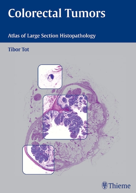
Correct diagnosis and staging are essential in determining the appropriate therapy of colorectal carcinoma, one of the most common malignancies in America and Europe. As medical science continues to develop rapidly, histopathology remains an essential part of diagnosis in most malignant diseases. In this era of interdisciplinary medicine, the role of pathology has expanded to provide images that easily correlate with endoscopic, radiological, or operative findings.
In this remarkable atlas, Tibor Tot presents colon pathology in large histological sections, with cross-sections of entire tumors in their anatomic environments and their circumferential surgical margins. These unique images form a guide to diagnosis, tumor typing and staging according to TNM criteria. They help to assess the completeness of a surgical excision and to understand the heterogeneity of colorectal carcinomas.
Features:
Cases illustrated in two-page spreads with clinical information, conventional histopathology, and large-section histology images enlarged to almost a full page.
Pathology seen in the context of surrounding tissues
The margins of malignant tumors visible in their entirety
Schematic guides to interpretation of the large-section images
Emphasis on diagnostic advantages of using large section technique
Technical guidelines for obtaining large-section histopathology specimens
This atlas is the result of seven years of studying almost 2,000 cases of colorectal carcinoma and other intestinal lesions and is highly recommended for pathologists, radiologists, surgeons, and oncologists alike.
Tibor Tot
| Sprache | englisch |
|---|---|
| Maße | 210 x 297 mm |
| Gewicht | 865 g |
| Themenwelt | Medizin / Pharmazie ► Medizinische Fachgebiete |
| Schlagworte | Colon • Colon / Kolon • Darm • HC/Medizin/Klinische Fächer • Histologie • Histopathology • Innere Medizin: Hämatologie/Onkologie • Krebs (Krankheit) • Krebs (Krankheit) / Karzinom • Radiologie • Studium Humanmedizin: 2. Studienabschnitt Pathologie • Tumor |
| ISBN-10 | 3-13-140591-0 / 3131405910 |
| ISBN-13 | 978-3-13-140591-3 / 9783131405913 |
| Zustand | Neuware |
| Haben Sie eine Frage zum Produkt? |
Mehr entdecken
aus dem Bereich
aus dem Bereich
Kompaktes Wissen, Sprachtraining und Simulationen für Mediziner
Buch | Softcover (2020)
Urban & Fischer in Elsevier (Verlag)
40,00 €


