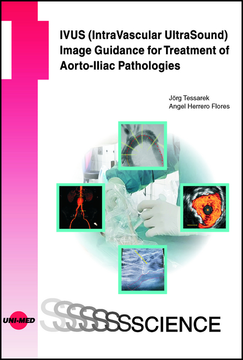
IVUS (IntraVascular UltraSound) Image Guidance for Treatment of Aorto-Iliac Pathologies
Part I1.Introduction121.1.US for access to target vessels121.2.US guidance for endovascular procedures131.3.Therapeutic use of US for endovascular reconstruction131.4.Why should we perform IVUS guidance for aortic and aorto-iliac procedures132.Radiation safety issues and risk awareness162.1.Revised radiation safety directives and guidelines 2020-2022162.2.Radiation safety items in the daily routine: staff training162.3.Radiation protection items in the daily routine172.4.Personalized radiation protection equipment182.5.Reduction of the individual radiation exposure202.5.1.Influence of radiation source position and individual behavior202.5.2.Influence of room geometry and procedural workflow on staff radiation exposure212.5.3.Projection angle and radiation safety: steady AP-projection for optimal radiation protection253.Iodinated Contrast Media (ICM) and procedure-related renal side effects303.1.CIN: Contrast-Induced Nephropathy313.1.1.Definition of CIN313.1.2.Pathophysiology of CIN313.1.3.Risk factors for CIN and prevention313.1.4.Differential diagnosis of CIN323.1.5.Strategies for CIN prevention333.1.6.IVUS for CIN prevention333.2.Acute kidney injury vs. CIN344.Software and hardware solutions for radiation dose / contrast media reduction and workflow improvement374.1.ClarityIQ(TM) and image noise reduction374.2.Image fusion solutions374.3.FORS (fiberoptic real shape imaging)384.4.Simulation-based endovascular (and open surgery) radiation protection training38Part II5.IVUS image guidance for endovascular procedures in respect to current radiation safety guidelines425.1.The revised ESVS radiation guidelines and the role of IVUS425.2.Decision making for IVUS use435.3.What is the current state of IVUS image guidance in the different vascular territories? Why use IVUS at all?445.3.1.IVUS for Percutaneous Coronary Interventions (PCI)445.3.2.IVUS for endovenous procedures445.3.3.IVUS for PAD455.3.4.Non-clinical aspects of IVUS: Cost effectiveness calculations for PVI and PCI455.3.5.IVUS and radiation/procedural safety466.Current technical state of IVUS image guidance506.1.Comparison of IVUS vs. angiographic imaging506.2.Catheter portfolio and technical specifications536.2.1.Technical basics of IV/US imaging536.2.2.IVUS catheter portfolio536.2.3.Technical specifications of IVUS catheters566.2.4.Imaging techniques and image information content596.2.4.1.ChromaFlo(TM)596.2.4.2.VH IVUS596.3.Current options for image processing606.3.1.Image processing with Intrasight 5S616.4.Standards of Procedure (SOP) for IVUS use636.4.1.SOP for the hybrid room646.4.2.SOP for the standard operating room (constricted room)656.5.Team challenge IVUS666.5.1.Team challenge IVUS catheter use666.5.2.Team challenge Pioneer Plus catheter use667.IVUS for EVAR697.1.Published IVUS data for TEVAR697.2.What are the key factors for advanced image guidance in TEVAR or aorto-iliac PAD procedures?707.3.Target vessel mapping and sealing zone definition by intraluminal cross-sectional imaging727.3.1.Parallaxes and IVUS737.4.SOPs for IVUS guidance during TEVAR / PAD747.4.1.EVAR-SOP in the local setting747.4.1.1.Vessel access747.4.1.2.Pullback maneuver with IVUS and image processing747.4.1.3.Mapping of the target vessels757.4.1.4.Catheter protection during graft deployment757.4.1.5.IVUS completion control757.4.1.6.Workspace setting for TEVAR / PAD757.5.IVUS image interpretation787.5.1.Image interpretation prior to infrarenal endograft placement797.5.2.Interpretation of IVUS images after endograft deployment (completion of IVUS pullback)837.5.3.Sealing length847.5.4.Endoleak detection with IVUS867.6.IVUS benefits in terms of prevention of early complications877.7.IVUS and radiation safety878.IVUS for TEVAR (for aneurysmal disease, aortic dissections, and other pathologies)938.1.Where and when IVUS is useful938.2.Procedural pathways and imaging properties for IVUS and angiography in different p
| Erscheinungsdatum | 11.01.2024 |
|---|---|
| Reihe/Serie | UNI-MED Science |
| Verlagsort | Bremen |
| Sprache | englisch |
| Maße | 176 x 246 mm |
| Gewicht | 412 g |
| Themenwelt | Medizinische Fachgebiete ► Innere Medizin ► Kardiologie / Angiologie |
| Schlagworte | Aortic Disease • Imaging • IVUS |
| ISBN-10 | 3-8374-1664-X / 383741664X |
| ISBN-13 | 978-3-8374-1664-0 / 9783837416640 |
| Zustand | Neuware |
| Haben Sie eine Frage zum Produkt? |
aus dem Bereich
