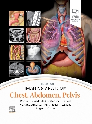
Imaging Anatomy: Chest, Abdomen, Pelvis
Churchill Livingstone (Verlag)
978-0-443-11800-5 (ISBN)
Contains nearly 2,800 print and online-only images, including all relevant imaging modalities, 3D reconstructions, and detailed, high-resolution medical drawings that together illustrate the fine points of imaging anatomy
Reflects new understandings of anatomy due to ongoing anatomic research as well as new, advanced imaging techniques
Offers new content on the anatomic basis for thoracic developmental abnormalities, anatomic variants of systemic and pulmonary vasculature, and the PI-RADS system and clinical implications of MR for prostate cancer
Contains new and updated images of the chest wall musculature with CT and MR examples; abdominal imaging best practices, including the application of body MR in the abdomen and pelvis; and the different modalities used for GU/GYN imaging, specifically retrograde urethrography and MR for specific disease diagnosis
Depicts common anatomic variants and covers the common pathological processes that manifest with alterations of normal anatomic landmarks
Features representative pathologic examples to highlight the effect of disease on human anatomy
Presents essential text in an easy-to-digest, bulleted format, enabling imaging specialists to find quick answers to anatomy questions encountered in daily practice
Includes an eBook version that enables you to access all text, figures, and references with the ability to search, customize your content, make notes and highlights, and have content read aloud
Dr. Siva Raman is a board-certified radiologist who gained his subspecialty expertise in thoracoabdominal imaging during a fellowship at Stanford University, and intern residency at UC Davis Medical Center. He attended Johns Hopkins University Medical School. Dr. Raman is a radiologist at Bay Imaging Consultants in Walnut Creek, California, with a subspecialty focus on thoracoabdominal imaging Melissa L. Rosado-de-Christenson, MD, FACR, FAAWR, is Attending Radiologist at the Division of Cardiothoracic Imaging in the Department of Medical Imaging for Banner - University Medical Group Tucson,. She is also Professor of Medical Imaging at University of Arizona College of Medicine - Tucson, in Tucson, Arizona Dr. Atif Zaheer has a specialty focus on CT, MR, and US of the abdomen and pelvis. He is Professor of Radiology, Oncology and Medicine at The Johns Hopkins University School of Medicine in Baltimore, Maryland. Dr. Santiago Martínez-Jiménez is with the Department of Radiology at Saint Luke's Hospital of Kansas City and is Professor of Radiology at the University of Missouri-Kansas City School of Medicine, in Kansas City, Missouri. He's a board-certified practicing radiologist who specializes in cardiothoracic radiology Dr. Ghaneh Fananapazir is a radiologist with specialization in abdominal radiology. He is Professor of Radiology at the University of California, Davis, in Sacramento, California. Fananapazir's clinical focus includes nonvascular interventional procedures, advanced US, CT and MR techniques, vascular imaging, and abdominal organ transplantation (kidney, liver, pancreas). In addition to his clinical duties, Dr. Fananapazir is passionate about medical education and research, particularly relating to kidney transplantation, and has authored textbooks and peer-reviewed research in highly impactful medical journals. Sherief H. Garrana, MD, is a clinical assistant professor of radiology with the Division of Thoracic Imaging in the Department of Radiology at NYU Langone Health in New York, New York Dr. Douglas Rogers is Associate Professor with the Department of Radiology and Imaging Services at University of Utah Health in Salt Lake City, Utah. Rogers is a board-certified Diagnostic Radiologist who specializes in abdominal imaging with a focus in gynecology. Dr. Bryan Foster is a body imaging radiologist with a subspecialty focus on oncologic, hepatobiliary, pancreatic, prostate, and small bowel imaging. Dr. Foster is the director of ultrasound at OHSU and performs complex image-guided biopsies; he also performs CEUS imaging and US elastography. Dr. Foster is skilled in all facets of imaging including radiography, fluoroscopy, ultrasound, CT, and MRI. His interests include oncologic, hepatobiliary, pancreatic, prostate, and small bowel imaging. Dr. Foster has recently started performing contrast-enhanced ultrasound imaging (as the only site in Oregon) and US elastography. He is one of two radiologists that perform MRI-guided prostate biopsies (also only site in Oregon). Recently he was selected as an Honored Educator for the Radiological Society of North America.
SECTION 1: CHEST 4 Chest Overview Melissa L. Rosado-de-Christenson, MD, FACR, FAAWR 44 Lung Development Melissa L. Rosado-de-Christenson, MD, FACR, FAAWR 66 Airway Structure Santiago Martínez-Jiménez, MD 88 Vascular Structure Santiago Martínez-Jiménez, MD 110 Interstitial Network Santiago Martínez-Jiménez, MD 122 Lungs Santiago Martínez-Jiménez, MD 154 Hila Melissa L. Rosado-de-Christenson, MD, FACR, FAAWR 186 Airways Melissa L. Rosado-de-Christenson, MD, FACR, FAAWR 212 Pulmonary Vessels Melissa L. Rosado-de-Christenson, MD, FACR, FAAWR 246 Systemic Vessels Santiago Martínez-Jiménez, MD and Melissa L. Rosado-de- Christenson, MD, FACR, FAAWR 284 Lymph Nodes and Lymphatics Sherief H. Garrana, MD 312 Pleura Santiago Martínez-Jiménez, MD 336 Mediastinum Melissa L. Rosado-de-Christenson, MD, FACR, FAAWR 370 Heart Melissa L. Rosado-de-Christenson, MD, FACR, FAAWR 414 Coronary Arteries and Cardiac Veins Sherief H. Garrana, MD 442 Pericardium Melissa L. Rosado-de-Christenson, MD, FACR, FAAWR 464 Chest Wall Sherief H. Garrana, MD SECTION 2: ABDOMEN 490 Embryology of Abdomen Atif Zaheer, MD, FSAR, Michael P. Federle, MD, FACR, and Siva P. Raman, MD 526 Abdominal Wall Atif Zaheer, MD, FSAR, Siva P. Raman, MD, and Michael P. Federle, MD, FACR 550 Diaphragm Atif Zaheer, MD, FSAR, Siva P. Raman, MD, and Michael P. Federle, MD, FACR 570 Peritoneal Cavity Atif Zaheer, MD, FSAR, Siva P. Raman, MD, and Michael P. Federle, MD, FACR 592 Vessels, Lymphatic System, and Nerves, Abdominal Atif Zaheer, MD, FSAR, Siva P. Raman, MD, and Michael P. Federle, MD, FACR 634 Esophagus Atif Zaheer, MD, FSAR, Siva P. Raman, MD, and Michael P. Federle, MD, FACR 650 Gastroduodenal Atif Zaheer, MD, FSAR, Siva P. Raman, MD, and Michael P. Federle, MD, FACR 678 Small Intestine Atif Zaheer, MD, FSAR, Siva P. Raman, MD, and Michael P. Federle, MD, FACR 708 Colon Atif Zaheer, MD, FSAR, Siva P. Raman, MD, and Michael P. Federle, MD, FACR 750 Spleen Siva P. Raman, MD 774 Liver Siva P. Raman, MD 820 Biliary System Siva P. Raman, MD 846 Pancreas Siva P. Raman, MD 876 Retroperitoneum Siva P. Raman, MD 902 Adrenal Siva P. Raman, MD 924 Kidney Siva P. Raman, MD 962 Ureter and Bladder Siva P. Raman, MD SECTION 3: PELVIS 988 Vessels, Lymphatic System, and Nerves, Pelvic Paula J. Woodward, MD, Akram M. Shaaban, MBBCh, and Ghaneh Fananapazir, MD, FSAR, FSRU, FSABI MALE 1016 Male Pelvic Wall and Floor Paula J. Woodward, MD, Akram M. Shaaban, MBBCh, and Ghaneh Fananapazir, MD, FSAR, FSRU, FSABI 1042 Testes and Scrotum Paula J. Woodward, MD, Akram M. Shaaban, MBBCh, and Ghaneh Fananapazir, MD, FSAR, FSRU, FSABI 1060 Prostate and Seminal Vesicles Bryan R. Foster, MD, Paula J. Woodward, MD, and Akram M. Shaaban, MBBCh 1078 Penis and Urethra Bryan R. Foster, MD, Paula J. Woodward, MD, and Akram M. Shaaban, MBBCh FEMALE 1094 Female Pelvic Floor Douglas Rogers, MD and Rania Farouk El Sayed, MD, PhD 1122 Uterus Douglas Rogers, MD 1148 Ovaries Douglas Rogers, MD
| Erscheinungsdatum | 26.10.2023 |
|---|---|
| Reihe/Serie | Imaging Anatomy |
| Verlagsort | London |
| Sprache | englisch |
| Maße | 216 x 276 mm |
| Gewicht | 3720 g |
| Themenwelt | Medizinische Fachgebiete ► Radiologie / Bildgebende Verfahren ► Radiologie |
| ISBN-10 | 0-443-11800-0 / 0443118000 |
| ISBN-13 | 978-0-443-11800-5 / 9780443118005 |
| Zustand | Neuware |
| Haben Sie eine Frage zum Produkt? |
aus dem Bereich


