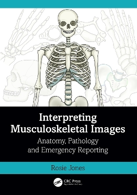
Interpreting Musculoskeletal Images
Anatomy, Pathology and Emergency Reporting
Seiten
2023
CRC Press (Verlag)
978-1-032-39891-4 (ISBN)
CRC Press (Verlag)
978-1-032-39891-4 (ISBN)
This visual manual with beautifully illustrated with schematic line diagrams, supplemented with radiographs and scans is an accessible guide to musculoskeletal image interpretation and reporting, including common trauma pathologies, arthropathies, mechanisms of injury and classification systems.
This visual manual is an accessible guide to musculoskeletal image interpretation and reporting, including common trauma pathologies, arthropathies, mechanisms of injury and classification systems. Beautifully illustrated with schematic line diagrams, supplemented with radiographs and scans, the content has been developed to enhance learning and understanding of both radiology and anatomy, and the relationship between them.
Key features:
Concise, yet highly informative
Large, high-quality illustrations supplement and enhance the written descriptions, with colour-coding for rapid matching of image to corresponding text
Relates imaging to underlying anatomy and pathology, aiding accurate interpretation
Carefully designed to support rapid access in the clinical setting and ideal also as a revision aid during examination preparation
The book delivers hands-on support to junior doctors, other emergency medicine personnel and practising radiographers for use in the clinical setting and is also ideal for students preparing for qualifying examinations in medicine and radiography.
Rosie Jones is a Radiographer at University of North Midlands NHS Trust, UK
Introduction
Upper limb
Lower limb
Pelvis and Hip
Spine
Face and Head
Paediatrics
Chest and Abdomen
Arthropathies
Bone lesions
Index
| Erscheinungsdatum | 01.09.2023 |
|---|---|
| Zusatzinfo | 237 Line drawings, color; 63 Halftones, black and white; 237 Illustrations, color; 63 Illustrations, black and white |
| Verlagsort | London |
| Sprache | englisch |
| Maße | 174 x 246 mm |
| Gewicht | 660 g |
| Themenwelt | Medizin / Pharmazie ► Allgemeines / Lexika |
| Medizin / Pharmazie ► Gesundheitsfachberufe ► MTA - Radiologie | |
| Medizin / Pharmazie ► Medizinische Fachgebiete ► Notfallmedizin | |
| Medizinische Fachgebiete ► Radiologie / Bildgebende Verfahren ► Radiologie | |
| Medizin / Pharmazie ► Physiotherapie / Ergotherapie ► Orthopädie | |
| Technik ► Medizintechnik | |
| Technik ► Umwelttechnik / Biotechnologie | |
| ISBN-10 | 1-032-39891-4 / 1032398914 |
| ISBN-13 | 978-1-032-39891-4 / 9781032398914 |
| Zustand | Neuware |
| Informationen gemäß Produktsicherheitsverordnung (GPSR) | |
| Haben Sie eine Frage zum Produkt? |
Mehr entdecken
aus dem Bereich
aus dem Bereich
Buch (2023)
Thieme (Verlag)
190,00 €


