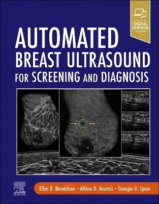
Automated Breast Ultrasound for Screening and Diagnosis
Elsevier - Health Sciences Division (Verlag)
978-0-323-55123-6 (ISBN)
- Noch nicht erschienen (ca. Januar 2026)
- Versandkostenfrei innerhalb Deutschlands
- Auch auf Rechnung
- Verfügbarkeit in der Filiale vor Ort prüfen
- Artikel merken
Volumetric automated breast ultrasound used to highlight the modality’s volumetric 3D qualities chosen from synonyms: ABUS, AUS, WHOBUS, or ABVS (tm) . Artifacts on Vo-AUS, especially shadowing--what causes them and how to resolve them . Primary screening, supplemental screening, and personalized screening for women with dense breasts-why and when to choose ultrasound . Performance of whole breast 3D volumetric automated breast ultrasound (Vo-AUS) for the supine and the prone patient with quality checklist . Artifacts on Vo-AUS, especially shadowing--what causes them and how to resolve them . Step-by-step protocol for efficient Interpretation of multiplanar screening Vo-AUS exams reviewed concurrently with AI-CAD for detection . Whole breast coronal plane: newly observed patterns for diagnosis . Breast anatomy and tissue composition depicted on whole breast Vo-AUS . Unique Atlas of Vo-AUS cases for BI-RADS assessment categories 1-6 . Vo-AUS chapters on calcifications, the postoperative breast, the male breast . Handheld US for the axilla and interventional guidance . Multimodality correlation with mammography, MRI, and tomosynthesis . Advances in ultrasound physics with current research and future directions . Includes 950 high-quality images - 900 radiographic and 50 color line drawings . Enhanced eBook version included with purchase. Your enhanced eBook allows you to access all of the text, figures, and references from the book on a variety of devices.
Ellen B. Mendelson, MD, MA, Professor Emeritus of Radiology, Feinberg School of Medicine, Northwestern University, Formerly Chief, Breast and Women's Imaging Division, Northwestern Medicine, Chicago, Illinois Athina Vourtsis, MD, PhD, Founder, Diagnostic Mammography Center, Athens, Greece Chief, Division of Breast Imaging, NorthShore University Health System, Clinical Associate Professor of Radiology, University of Chicago, Chicago, Illinois
Foreword
Preface
Section 1. Introduction: Evolution of Breast Ultrasound
1. A Brief History of Automated Whole Breast Ultrasound
Section 2. Automated Volumetric Whole Breast Ultrasound (Vo-AUS): Performance and Interpretation
2. Ultrasound for Breast Cancer Screening
3. Role of AI in breast ultrasound
4. BI-RADS for Ultrasound
5. Ultrasound Physics and Image Generation
6. Performing Automated Volumetric Breast Ultrasound for the Supine Patient
7. Interpreting Automated Volumetric Breast Ultrasound for the Supine Patient
8. Performing and Interpreting Automated Volumetric Breast Ultrasound for the Prone Patient
Section 3. Automated Atlas of Cases
9. Normal Anatomy and Tissue Composition: BI-RADS 1
10. Benign Findings: BI-RADS 2
11. Probably Benign and Low Suspicion Assessments: BI-RADS 3 and 4a
12. Vo-AUS Detection and Diagnosis of Calcifications
13. Suspicious Assessments: BI-RADS 4b and 4c
14. Highly Suggestive of Malignancy: BI-RADS 5 Cases
15. Known Carcinoma: BI-RADS 6
16. Lymph Nodes
17. Post-operative changes
18. Male Breast
19. Artifacts of Vo-AUS
20. Handheld Ultrasound-Guided Interventions for Vo-AUS Detected Findings
21. Prone Automated Breast Ultrasound Case Studies
Section 4. Implementation and Integration of Vo-AUS in Practice
22. Implementation of Automated Breast Ultrasound
23. Legislative and Economic Considerations
Section 5 Conclusion: Future of Breast Ultrasound
24. Current Research and New Technologies
| Erscheint lt. Verlag | 2.1.2026 |
|---|---|
| Zusatzinfo | Approx. 1000 illustrations (100 in full color); Illustrations |
| Verlagsort | Philadelphia |
| Sprache | englisch |
| Maße | 216 x 276 mm |
| Themenwelt | Medizinische Fachgebiete ► Radiologie / Bildgebende Verfahren ► Radiologie |
| ISBN-10 | 0-323-55123-8 / 0323551238 |
| ISBN-13 | 978-0-323-55123-6 / 9780323551236 |
| Zustand | Neuware |
| Haben Sie eine Frage zum Produkt? |
aus dem Bereich


