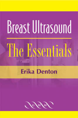
Breast Ultrasound: The Essentials
Seiten
2005
Greenwich Medical Media Ltd (Verlag)
978-1-84110-176-7 (ISBN)
Greenwich Medical Media Ltd (Verlag)
978-1-84110-176-7 (ISBN)
- Titel wird leider nicht erscheinen
- Artikel merken
This superbly illustrated text begins with an introduction to the equipment used and techniques needed to successfully image breast tissue, and then presents about 60 'classic', fully illustrated cases that might be encountered by the radiologist or sonographer, including a few rarer cases or cases that could easily be misinterpreted.
Breast ultrasound is an important diagnostic tool in the fight against breast cancer, especially in young (pre-menopausal) women, where it can have a greater sensitivity than mammography. Even in older women, ultrasound is becoming increasingly used as an adjunct to mammography, and as the imaging modality of choice when undertaking image-guided core biopsies or fine needle aspiration from suspect breast tissue. This superbly illustrated text begins with an introduction to the equipment used and techniques needed to successfully image breast tissue, and then goes on to present about 60 'classic', fully illustrated cases that might be encountered by the radiologist or sonographer, including a few rarer cases or cases that could easily be misinterpreted. In each case, clear images are provided, and the explanatory text takes the reader through the key features of interest and the important diagnostic points.
Breast ultrasound is an important diagnostic tool in the fight against breast cancer, especially in young (pre-menopausal) women, where it can have a greater sensitivity than mammography. Even in older women, ultrasound is becoming increasingly used as an adjunct to mammography, and as the imaging modality of choice when undertaking image-guided core biopsies or fine needle aspiration from suspect breast tissue. This superbly illustrated text begins with an introduction to the equipment used and techniques needed to successfully image breast tissue, and then goes on to present about 60 'classic', fully illustrated cases that might be encountered by the radiologist or sonographer, including a few rarer cases or cases that could easily be misinterpreted. In each case, clear images are provided, and the explanatory text takes the reader through the key features of interest and the important diagnostic points.
| Erscheint lt. Verlag | 31.8.2005 |
|---|---|
| Zusatzinfo | Worked examples or Exercises; Worked examples or Exercises |
| Verlagsort | Cambridge |
| Sprache | englisch |
| Themenwelt | Medizin / Pharmazie ► Medizinische Fachgebiete ► Onkologie |
| Medizinische Fachgebiete ► Radiologie / Bildgebende Verfahren ► Sonographie / Echokardiographie | |
| ISBN-10 | 1-84110-176-1 / 1841101761 |
| ISBN-13 | 978-1-84110-176-7 / 9781841101767 |
| Zustand | Neuware |
| Haben Sie eine Frage zum Produkt? |
Mehr entdecken
aus dem Bereich
aus dem Bereich
Begleitbuch für Sonografiekurse, Klinik und Praxis
Buch | Softcover (2023)
Urban & Fischer in Elsevier (Verlag)
27,00 €
Organbezogene Darstellung von Grund- und Aufbaukurs sowie …
Buch | Hardcover (2020)
Deutscher Ärzteverlag
99,99 €


