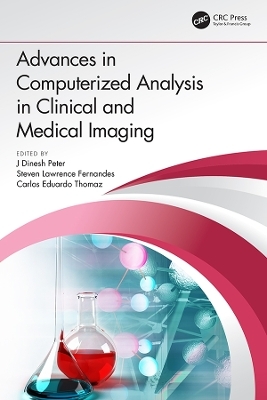
Advances in Computerized Analysis in Clinical and Medical Imaging
CRC Press (Verlag)
978-1-138-33329-1 (ISBN)
Advances in Computerized Analysis in Clinical and Medical Imaging book is devoted for spreading of knowledge through the publication of scholarly research, primarily in the fields of clinical & medical imaging. The types of chapters consented include those that cover the development and implementation of algorithms and strategies based on the use of geometrical, statistical, physical, functional to solve the following types of problems, using medical image datasets: visualization, feature extraction, segmentation, image-guided surgery, representation of pictorial data, statistical shape analysis, computational physiology and telemedicine with medical images.
This book highlights annotations for all the medical and clinical imaging researchers’ a fundamental advances of clinical and medical image analysis techniques. This book will be a good source for all the medical imaging and clinical research professionals, outstanding scientists, and educators from all around the world for network of knowledge sharing. This book will comprise high quality disseminations of new ideas, technology focus, research results and discussions on the evolution of Clinical and Medical image analysis techniques for the benefit of both scientific and industrial developments.
Features:
Research aspects in clinical and medical image processing
Human Computer Interaction and interface in imaging diagnostics
Intelligent Imaging Systems for effective analysis using machine learning algorithms
Clinical and Scientific Evaluation of Imaging Studies
Computer-aided disease detection and diagnosis
Clinical evaluations of new technologies
Mobility and assistive devices for challenged and elderly people
This book serves as a reference book for researchers and doctoral students in the clinical and medical imaging domain including radiologists. Industries that manufacture imaging modality systems and develop optical systems would be especially interested in the challenges and solutions provided in the book. Professionals and practitioners in the medical and clinical imaging may be benefited directly from authors’ experiences.
J. Dinesh Peter is currently working as Associate Professor, Department of Computer Science and Engineering at Karunya Institute of Technology & Sciences, Coimbatore. Prior to this, he was a full time research scholar at National Institute of Technology, Calicut, India, from where he received his PhD in computer science and engineering. His research focus includes Big-data, image processing and computer vision. He has several publications in various reputed international journals and conference papers which are widely referred to. He is a member of IEEE, MICCAI, Computer Society of India and Institution of Engineers India and has served as session chairs and delivered plenary speeches for various international conferences and workshops. He has conducted many international conferences and been as editor for Springer proceedings and many special issues in journals. Steven Lawrence Fernandes is currently working as a postdoctoral researcher in the area of deep learning under the guidance of Professor Sumit Kumar Jha at The University of Central Florida, USA. He also has postdoctoral research experience working at The University of Alabama at Birmingham, USA. He has his Ph.D. in Computer Vision and Machine Learning from Karunya Institute of Technology & Sciences, Coimbatore, Tamil Nadu. His Ph.D work "Match Composite Sketch with Drone Images" has received patent notification (Patent Application Number: 2983/CHE/2015) from Government of India, Controller General of Patents, Designs & Trade Marks. He has received the prestigious US award from Society for Design and Process Science for his outstanding service contributions in the year 2017 and Young Scientist Award by Vision Group on Science and Technology, Government of Karnataka, India in the year 2014. He also received Research Grant from University of Houston Downtown, USA and The Institution of Engineers (India), Kolkata. He has collaborated with various Scientists, Professors, Researchers and jointly published more than 50 Research Articles which are in Science Citation Indexed (SCI) Journals. Carlos E. Thomaz holds a degree in Electronic Engineering from the Pontifical Catholic University of Rio de Janeiro (1993), a Master's degree in Electrical Engineering from the Pontifical Catholic University of Rio de Janeiro (1999), a PhD and a postdoctoral degree in Computer Science - Imperial College London (2005). He is a full professor at FEI's University Center. He has experience in the area of Computer Science, with emphasis on Pattern Recognition in Statistics, working mainly in the following subjects: Computational Vision, Computation in Medical Images and Biometrics.
1. A New Biomarker for Alzheimer’s Based on the Hippocampus Image Through the Evaluation of the Surface Charge Distribution 2. Independent Vector Analysis of Non-Negative Image Mixture Model for Clinical Image Separation 3. Rationalizing of Morphological Renal Parameters and eGFR for Chronic Kidney Disease Detection 4. Human Computer Interface for Neurodegenerative Patients Using Machine Learning Algorithms 5. Smart Mobility System for Physically Challenged People 6. DHS: The Cognitive Companion for Assisted Living of the Elderly 7. Raspberry Pi Based Cancer Cell Detection Using Segmentation Algorithm 8. An AAC Communication Device for Patients with Total Paralysis 9. Case Studies on Medical Diagnosis Using Soft Computing Techniques 10. Alzheimer’s Disease Classification Using Machine Learning Algorithms 11. Fetal Standard Plane Detection in Freehand Ultrasound Using Multi Layered Extreme Learning Machine 12. Earlier Prediction of Cardiovascular Disease Using IoT and Deep Learning Approaches 13. Analysis of Heart Disease Prediction Using Various Machine Learning Techniques 14. Computer-Aided Detection of Breast Cancer on Mammograms: Extreme Learning Machine Neural Network Approach 15. Deep Learning Segmentation Techniques for Checking the Anomalies of White Matter Hyperintensities in Alzheimer’s Patients 16. Investigations on Stabilization and Compression of Medical Videos 17. An Automated Hybrid Methodology Using Firefly Based Fuzzy Clustering for Demarcation of Tissue and Tumor Region in Magnetic Resonance Brain Images 18. A Risk Assessment Model for Alzheimer’s Disease Using Fuzzy Cognitive Map 19. Comparative Analysis of Texture Patterns for the Detection of Breast Cancer Using Mammogram Images 20. Analysis of Various Color Models for Endoscopic Images 21. Adaptive Fractal Image Coding Using Differential Scheme for Compressing Medical Images
| Erscheinungsdatum | 13.05.2019 |
|---|---|
| Zusatzinfo | 53 Tables, black and white; 178 Illustrations, black and white |
| Verlagsort | London |
| Sprache | englisch |
| Maße | 178 x 254 mm |
| Gewicht | 657 g |
| Themenwelt | Informatik ► Theorie / Studium ► Künstliche Intelligenz / Robotik |
| Medizin / Pharmazie ► Medizinische Fachgebiete ► Radiologie / Bildgebende Verfahren | |
| Studium ► Querschnittsbereiche ► Prävention / Gesundheitsförderung | |
| ISBN-10 | 1-138-33329-8 / 1138333298 |
| ISBN-13 | 978-1-138-33329-1 / 9781138333291 |
| Zustand | Neuware |
| Haben Sie eine Frage zum Produkt? |
aus dem Bereich


