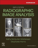
Workbook for Radiographic Image Analysis
Seiten
2019
|
5th edition
Saunders (Verlag)
978-0-323-54463-4 (ISBN)
Saunders (Verlag)
978-0-323-54463-4 (ISBN)
- Titel erscheint in neuer Auflage
- Artikel merken
Zu diesem Artikel existiert eine Nachauflage
Get all the tools you need to hone your imaging and evaluation skills with Kathy Martensen's Workbook for Radiographic Image Analysis, 5th Edition. This complete workbook offers ample opportunities to practice and apply information from the main Radiographic Image Analysis text via study questions for each procedure, positioning and technique exercises, and additional suboptimal images to identify. This new workbook edition features updated content that reflects the latest ARRT guidelines plus additional images not found in the main text. Workbook users can easily check your work in the answer key found in the back of the book.
Study questions reinforce text material and prepare you for certification.
Incorrectly positioned images with questions ensure you understand what features need to be visible in an image and how to adjust when the images are poor.
Additional images not included in the main text offer additional practice with identifying poor quality images and recognizing how they are produced.
Positioning and technique exercises prepare you for success in radiography practice.
NEW! Updated content reflects the latest ARRT guidelines.
NEW! Additional images offer further visual guidance to help you better critique and correct positioning errors.
NEW! More robust digital halftones across images paint a clearer picture of proper technique.
1. Guidelines for Image Analysis
2. Visibility of Details
3. Image Analysis of the Chest and Abdomen
4. Image Analysis of the Upper Extremity
5. Image Analysis of the Shoulder
6. Image Analysis of the Lower Extremity
7. Image Analysis of the Hip and Pelvis
8. Image Analysis of the Cervical and Thoracic Vertebrae
9. Image Analysis of the Lumbar Vertebrae, Sacrum, and Coccyx
10. Image Analysis of the Sternum and Ribs
11. Image Analysis of the Cranium
12. Image Analysis of the Digestive System
| Erscheinungsdatum | 27.02.2019 |
|---|---|
| Zusatzinfo | 605 illustrations; Illustrations |
| Verlagsort | Philadelphia |
| Sprache | englisch |
| Maße | 216 x 276 mm |
| Gewicht | 970 g |
| Themenwelt | Medizin / Pharmazie ► Gesundheitsfachberufe ► MTA - Radiologie |
| Medizinische Fachgebiete ► Radiologie / Bildgebende Verfahren ► Sonographie / Echokardiographie | |
| ISBN-10 | 0-323-54463-0 / 0323544630 |
| ISBN-13 | 978-0-323-54463-4 / 9780323544634 |
| Zustand | Neuware |
| Haben Sie eine Frage zum Produkt? |
Mehr entdecken
aus dem Bereich
aus dem Bereich
Begleitbuch für Sonografiekurse, Klinik und Praxis
Buch | Softcover (2023)
Urban & Fischer in Elsevier (Verlag)
27,00 €
Organbezogene Darstellung von Grund- und Aufbaukurs sowie …
Buch | Hardcover (2020)
Deutscher Ärzteverlag
99,99 €


