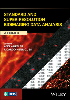
Standard and Super-Resolution Bioimaging Data Analysis
John Wiley & Sons Inc (Verlag)
978-1-119-09690-0 (ISBN)
- Titel z.Zt. nicht lieferbar
- Versandkostenfrei innerhalb Deutschlands
- Auch auf Rechnung
- Verfügbarkeit in der Filiale vor Ort prüfen
- Artikel merken
Standard and Super-Resolution Bioimaging Data Analysis gets newcomers to bioimage data analysis quickly up to speed on the mathematics, statistics, computing hardware and acquisition technologies required to correctly process and document data.
The past quarter century has seen remarkable progress in the field of light microscopy for biomedical science, with new imaging technologies coming on the market at an almost annual basis. Most of the data generated by these systems is image-based, and there is a significant increase in the content and throughput of these imaging systems. This, in turn, has resulted in a shift in the literature on biomedical research from descriptive to highly-quantitative. Standard and Super-Resolution Bioimaging Data Analysis satisfies the demand among students and research scientists for introductory guides to the tools for parsing and processing image data. Extremely well illustrated and including numerous examples, it clearly and accessibly explains what image data is and how to process and document it, as well as the current resources and standards in the field.
A comprehensive guide to the tools for parsing and processing image data and the resources and industry standards for the biological and biomedical sciences
Takes a practical approach to image analysis to assist scientists in ensuring scientific data are robust and reliable
Covers fundamental principles in such a way as to give beginners a sound scientific base upon which to build
Ideally suited for advanced students having only limited knowledge of the mathematics, statistics and computing required for image data analysis
An entry-level text written for students and practitioners in the bioscience community, Standard and Super-Resolution Bioimaging Data Analysis de-mythologises the vast array of image analysis modalities which have come online over the past decade while schooling beginners in bioimaging principles, mathematics, technologies and standards.
ANN WHEELER, PhD, is Head of the Advanced Imaging Resource at the MRC Institute of Genetics and Molecular Medicine, University of Edinburgh, UK. RICARDO HENRIQUES, PhD, is Head of the Quantitative Imaging and NanoBioPhysics research group at the MRC Laboratory for Molecular Cell Biology, University College London, UK.
List of Contributors xi
Foreword xiii
1 Digital Microscopy: Nature to Numbers 1
Ann Wheeler
1.1 Acquisition 4
1.1.1 First Principles: How Can Images Be Quantified? 4
1.1.2 Representing Images as a Numerical Matrix Using a Scientific Camera 6
1.1.3 Controlling Pixel Size in Cameras 8
1.2 Initialisation 11
1.2.1 The Sample 12
1.2.2 Pre]Processing 12
1.2.3 Denoising 12
1.2.4 Filtering Images 14
1.2.5 Deconvolution 16
1.2.6 Registration and Calibration 19
1.3 Measurement 21
1.4 Interpretation 23
1.5 References 29
2 Quantification of Image Data 31
Jean]Yves Tinevez
2.1 Making Sense of Images 31
2.1.1 The Magritte Pipe 31
2.1.2 Quantification of Image Data Via Computers 33
2.2 Quantifiable Information 35
2.2.1 Measuring and Comparing Intensities 35
2.2.2 Quantifying Shape 36
2.2.3 Spatial Arrangement of Objects 41
2.3 Wrapping Up 45
2.4 References 46
3 Segmentation in Bioimaging 47
Jean]Yves Tinevez
3.1 Segmentation and Information Condensation 47
3.1.1 A Priori Knowledge 48
3.1.2 An Intuitive Approach 49
3.1.3 A Strategic Approach 51
3.2 Extracting Objects 52
3.2.1 Detecting and Counting Objects 52
3.2.2 Automated Segmentation of Objects 60
3.3 Wrapping Up 74
3.4 References 79
4 Measuring Molecular Dynamics and Interactions by Förster Resonance Energy Transfer (FRET) 83
Aliaksandr Halavatyi and Stefan Terjung
4.1 FRET]Based Techniques 83
4.1.1 Ratiometric Imaging 84
4.1.2 Acceptor Photobleaching 85
4.1.3 Other FRET Measurement Techniques 85
4.1.4 Alternative Methods to Measure Interactions 87
4.2 Experimental Design 89
4.2.1 Ratiometric Imaging of FRET]Based Sensors 90
4.2.2 Acceptor Photobleaching 91
4.3 FRET Data Analysis 92
4.3.1 Ratiometric Imaging 92
4.3.2 Acceptor Photobleaching 93
4.3.3 Data Averaging and Statistical Analysis 93
4.4 Computational Aspects of Data Processing 94
4.4.1 Software Tools 94
4.4.2 FRET Data Analysis with Fiji 94
4.5 Concluding Remarks 95
4.6 References 96
5 FRAP and Other Photoperturbation Techniques 99
Aliaksandr Halavatyi and Stefan Terjung
5.1 Photoperturbation Techniques in Cell Biology 99
5.1.1 Scientific Principles Underpinning FRAP 100
5.1.2 Other Photoperturbation Techniques 103
5.2 FRAP Experiments 106
5.2.1 Selecting Fluorescent Tags 107
5.2.2 Optimisation of FRAP Experiments 107
5.2.3 Storage of Experimental Data 109
5.3 FRAP Data Analysis 109
5.3.1 Quantification of FRAP Intensities 112
5.3.2 Normalisation 113
5.3.3 In Silico Modelling of FRAP Data 115
5.3.4 Fitting Recovery Curves 120
5.3.5 Evaluating the Quality of FRAP Data and Analysis Results 121
5.3.6 Data Averaging and Statistical Analysis 122
5.3.7 Software for FRAP Data Processing 123
5.4 Procedures for Quantitative FRAP Analysis with Freeware Software Tools 127
5.4.1 Quantification of Intensity Traces with Fiji 127
5.4.2 Processing FRAP Recovery Curves with FRAPAnalyser 128
5.5 Notes 130
5.6 Concluding Remarks 131
5.7 References 132
5A Case Study: Analysing COPII Turnover During ER Exit 135
5A.1 Quantitative FRAP Analysis of ER-Exit Sites 135
5A.2 Mechanistic Insight into COPII Coat Kinetics with FRAP 138
5A.3 Automated FRAP at ERESs 140
5A.4 References 141
6 Co]Localisation and Correlation in Fluorescence Microscopy Data 143
Dylan Owen, George Ashdown, Juliette Griffié and Michael Shannon
6.1 Introduction 143
6.2 Co]Localisation for Conventional Microscopy Images 145
6.2.1 Co]Localisation in Super]Resolution Localisation Microscopy 151
6.2.2 Fluorescence Correlation Spectroscopy 156
6.2.3 Image Correlation Spectroscopy 161
6.3 Conclusion 164
6.4 Acknowledgments 165
6.5 References 165
7 Live Cell Imaging: Tracking Cell Movement 173
Mario De Piano, Gareth E. Jones and Claire M. Wells
7.1 Introduction 173
7.2 Setting up a Movie for Time]Lapse Imaging 174
7.3 Overview of Automated and Manual Cell Tracking Software 175
7.3.1 Automatic Tracking 176
7.3.2 Manual Tracking 180
7.3.3 Comparison Between Automated and Manual Tracking 181
7.4 Instructions for Using ImageJ Tracking 184
7.5 Post]Tracking Analysis Using the Dunn Mathematica Software 189
7.6 Summary and Future Direction 198
7.7 References 198
8 Super]Resolution Data Analysis 201
Debora Keller, Nicolas Olivier, Thomas Pengo and Graeme Ball
8.1 Introduction to Super]Resolution Microscopy 201
8.2 Processing Structured Illumination Microscopy Data 202
8.2.1 SIM Reconstruction Theory 203
8.2.2 Parameter Fitting and Corrections 204
8.2.3 SIM Quality Control 205
8.2.4 Checking System Calibration 205
8.2.5 Checking Raw Data 205
8.2.6 Checking Reconstructed Data 208
8.2.7 SIM Data Analysis 208
8.3 Quantifying Single Molecule Localisation Microscopy Data 210
8.3.1 SMLMS Pre]Processing 210
8.3.2 Localisation: Finding Molecule Positions 210
8.3.3 Fitting Molecules 210
8.3.4 Problem of Multiple Emissions Per Molecule 212
8.3.5 Sieving and Quality Control and Drift Correction 213
8.3.6 How Far Can I Trust the SMLM Data? 218
8.4 Reconstruction Summary 220
8.5 Image Analysis on Localisation Data 220
8.5.1 Cluster Analysis 221
8.5.2 Stoichiometry and Counting 222
8.5.3 Fitting and Particle Averaging 223
8.5.4 Tracing 223
8.6 Summary and Available Tools 223
8.7 References 224
9 Big Data and Bio]Image Informatics: A Review of Software Technologies Available for Quantifying Large Datasets in Light]Microscopy 227
Ahmed Fetit
9.1 Introduction 227
9.2 What Is Big Data Anyway? 228
9.3 The Open]Source Bioimage Informatics Community 231
9.3.1 ImageJ for Small]Scale Projects 231
9.3.2 CellProfiler, Large]Scale Projects and the Need for Complex Infrastructure 235
9.3.3 Technical Notes – Setting Up CellProfiler for Use on a Linux HPC 238
9.3.4 Icy, Towards Reproducible Image Informatics 242
9.4 Commercial Solutions for Bioimage Informatics 243
9.4.1 Imaris Bitplane 243
9.4.2 Definiens and Using Machine]Learning on Complex Datasets 244
9.5 Summary 247
9.6 Acknowledgments 247
9.7 References 248
10 Presenting and Storing Data for Publication 249
Ann Wheeler and Sébastien Besson
10.1 How to Make Scientific Figures 249
10.1.1 General Guidelines for Making Any Microscopy Figure 250
10.1.2 Do’s and Don’ts: Preparation of Figures for Publication 251
10.1.3 Restoration, Revelation or Manipulation 253
10.2 Presenting, Documenting and Storing Bioimage Data 256
10.2.1 Metadata Matters 257
10.2.2 The Open Microscopy Project 258
10.2.3 OME and Bio]Formats, Supporting Interoperability in Bioimaging Data 259
10.2.4 Long]Term Data Storage 260
10.2.5 USB Drives Friend or Foe? 262
10.2.6 Beyond the (USB) Drive Limit 262
10.2.7 Servers and Storage Area Networks 263
10.2.8 OMERO Scalable Data Management for Biologists 265
10.3 Summary 267
10.4 References 268
11 Epilogue: A Framework for Bioimage Analysis 269
Kota Miura and Sébastien Tosi
11.1 Workflows for Bioimage Analysis 270
11.1.1 Components 270
11.1.2 Workflows 272
11.1.3 Types of Workflows 273
11.1.4 Types of Component 276
11.2 Resources for Designing Workflows and Supporting Bioimage Analysis 277
11.2.1 A Brief History 278
11.2.2 A Network for Bioimage Analysis 279
11.2.3 Additional Textbooks 279
11.2.4 Training Schools 280
11.2.5 Database of Components and Workflows 280
11.2.6 Benchmarking Platform 282
11.3 Conclusion 282
11.4 References 283
Index 285
| Erscheinungsdatum | 16.03.2018 |
|---|---|
| Reihe/Serie | RMS - Royal Microscopical Society |
| Verlagsort | New York |
| Sprache | englisch |
| Maße | 155 x 231 mm |
| Gewicht | 635 g |
| Themenwelt | Medizin / Pharmazie ► Medizinische Fachgebiete ► Radiologie / Bildgebende Verfahren |
| Medizin / Pharmazie ► Physiotherapie / Ergotherapie ► Orthopädie | |
| Naturwissenschaften ► Biologie | |
| Naturwissenschaften ► Chemie | |
| Naturwissenschaften ► Physik / Astronomie | |
| Technik ► Maschinenbau | |
| Technik ► Medizintechnik | |
| ISBN-10 | 1-119-09690-1 / 1119096901 |
| ISBN-13 | 978-1-119-09690-0 / 9781119096900 |
| Zustand | Neuware |
| Informationen gemäß Produktsicherheitsverordnung (GPSR) | |
| Haben Sie eine Frage zum Produkt? |
aus dem Bereich


