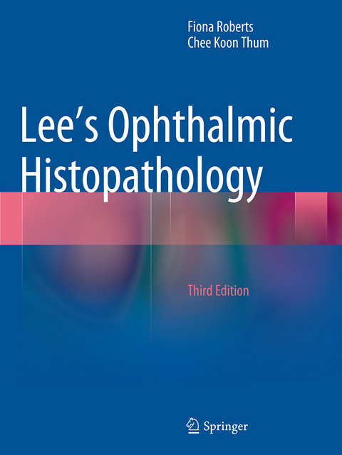
Lee's Ophthalmic Histopathology
Seiten
2016
|
Softcover reprint of the original 3rd ed. 2014
Springer London Ltd (Verlag)
978-1-4471-7117-1 (ISBN)
Springer London Ltd (Verlag)
978-1-4471-7117-1 (ISBN)
This well-illustrated, practical text emphasizes recent advances, particularly in the molecular basis of disease and in the diagnosis and classification of tumors. Specimen-based chapter layout gives a quick lead into the handling of this type of specimen.
Completely revised and updated, this well-illustrated and practically-oriented text has retained its general layout and style and division into specimen-type based chapters. The visual image remains key to explaining the pathological processes and this is facilitated by full colour photography throughout the text. The book emphasizes pertinent recent advances which augment the morphological study of disease. There is updated information on clinically important aspects, immunohistochemistry, tumour cytogenetics and molecular biology.
Illustrations include macro specimens, microscopic specimens and illustrations of additional techniques - immunohistochemistry, molecular analyses and electron microscopy - to help both the pathologist and ophthalmologist understand the process that a specimen must go through prior to producing a report and how these various techniques help to refine the diagnosis.
The third edition of Lee’s Ophthalmic Histopathology is an invaluable reference source for ophthalmic pathologists, general pathologists and ophthalmologists.
Fiona Roberts, Southern General Hospital, Glasgow, UK Chee Koon Thum, Western General Hospital, Edinburgh, UK
Examination of the Globe: Technical Aspects.- The Traumatised Eye.- “Absolute Glaucoma”.- Retinal Vascular Disease.- Intraocular Tumours.- Ocular Inflammation.- Treatment of Retinal Detachment.- The Malformed Eye.- “Autopsy Eye”: the Eye in Systemic Disease.- Biopsy of the Eyelid, the Lacrimal Sac, and the Temporal Artery.- The Conjunctival Biopsy.- The Orbit: Biopsy, Excision Biopsy, and Exenteration Specimens.- The Corneal Disc.- Lens.
| Erscheinungsdatum | 14.10.2016 |
|---|---|
| Zusatzinfo | 535 Illustrations, color; 39 Illustrations, black and white; XXVII, 466 p. 574 illus., 535 illus. in color. |
| Verlagsort | England |
| Sprache | englisch |
| Maße | 210 x 279 mm |
| Themenwelt | Medizin / Pharmazie ► Medizinische Fachgebiete ► Augenheilkunde |
| Studium ► 2. Studienabschnitt (Klinik) ► Pathologie | |
| ISBN-10 | 1-4471-7117-9 / 1447171179 |
| ISBN-13 | 978-1-4471-7117-1 / 9781447171171 |
| Zustand | Neuware |
| Haben Sie eine Frage zum Produkt? |
Mehr entdecken
aus dem Bereich
aus dem Bereich


