Ultrasound
Oxford University Press Inc (Verlag)
978-0-19-998810-5 (ISBN)
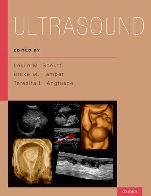
In 187 cases that feature over 1,700 high-quality images, Ultrasound provides a succinct review of clinically relevant cases covering the full range of clinical problems and diagnoses in this subspecialty of radiology. Pathologies are grouped into Gynecologic, Obstetrical, Abdominal, Small Parts, and Vascular sections. The volume follows the easy-to-use format of question and answer in which the patient history is provided on the first page of the case, and radiologic findings, differential diagnoses, teaching points, and next steps in management, followed by suggestions for further reading. Ultrasound is an essential resource for radiology residents and practicing radiologists alike.
Leslie M. Scoutt is a Professor of Diagnostic Radiology and Vascular Surgery, Vice Chair of Education, Department of Diagnostic Radiology, Chief of the Ultrasound Section, Medical Director of the Non-Invasive Vascular Laboratory, and the Associate Program Director in the Department of Radiology and Biomedical Imaging at the Yale University School of Medicine in New Haven, Connecticut. Ulrike M. Hamper is a Professor of Radiology, Urology, and Pathology and the Director of the Ultrasound Division in the Russell H. Morgan Department of Radiology and Radiological Science at the Johns Hopkins University School of Medicine in Baltimore, Maryland Teresita L. Angtuaco is the Director of the Division of Body Imaging and the Chief of Ultrasound in the Department of Radiology at the University of Arkansas for Medical Sciences in Little Rock, Arkansas
Section I. Gynecology
1. Hydrosalpinx
2. Bilateral Tubo-Ovarian Abscess
3. Postmenopausal Ovarian Cyst
4. Mucinous Ovarian Cystadenoma
5. Serous Cystadenocarcinoma of the Ovary
6. Bilateral Krukenberg Tumors
7. Adenomyosis of the Uterus
8. Lipoleiomyoma
9. Endometrial Cancer
10. Uterine Arteriovenous Malformation
11. Hemorrhagic Cyst
12. Endometrioma
13. Dermoid Cyst
14. Peritoneal Inclusion Cyst
15. Ovarian Fibrothecoma
16. Ovarian Torsion
17. Endometritis
18. Misplaced IUD
19. Endometrial Polyp
20. Endometrial Hyperplasia
21. Pedunculated Subserosal Leiomyoma
22. Septate Uterus
23. Cervical Cancer
Section II. First Trimester
24. Intradecidual Sign
25. First Trimester Perigestational Hemorrhage
26. Anembryonic Gestation
27. Twin Chorionicity
28. Open Rhombencephalon
29. Physiologic Gut Herniation
30. Tubal Ectopic Pregnancy
31. Interstitial Ectopic Pregnancy
32. Ovarian Hyperstimulation Syndrome with Heterotopic Pregnancies
33. Pregnancy in C-Section Scar
34. Incomplete Abortion
35. Coexistent Molar Gestation
36. Nuchal Translucency
Section III. Second Trimester
37. Cervical Incompetence
38. Placenta Previa
39. Placenta Percreta
40. Placental Abruption
41. Hydranencephaly
42. Alobar Holoprosencephaly
43. Vein of Galen Malformation
44. Dandy Walker Malformation
45. Agenesis of the Corpus Callosum
46. Chiari II Malformation
47. Cleft Lip and Palate
48. Congenital Pulmonary Airway Malformation (CPAM)
49. Hypoplastic Left Heart Syndrome
50. Transposition of the Great Arteries
51. Omphalocele
52. Gastroschisis
53. Congenital Diaphragmatic Hernia
54. Esophageal Atresia
55. Duodenal Atresia
56. Multicystic Dysplastic Kidney
57. Ureteropelvic Junction Obstruction
58. Autosomal Recessive Polycystic Kidney Disease
59. Posterior Urethral Valves
60. Renal Agenesis
61. Beckwith Wiedemann Syndrome
62. Trisomy 21
63. Trisomy 18
64. Trisomy 13
65. Twin Transfusion Syndrome
66. Meconium Pseudocyst
67. Osteogenesis Imperfecta Type II
Section IV. Abdomen
68. Liver Abscess
69. Focal Nodular Hyperplasia
70. Gallbladder Wall Thickening Due to Hepatitis
71. Adenomyomatosis
72. Caroli Disease
73. Acute Pancreatitis
74. Appendicitis
75. Intussusception
76. Prostatic Abscess
77. Renal Angiomyelolipoma
78. Horseshoe Kidney
79. Crossed Fused Renal Ectopia
80. Emphysematous Pyelonephritis
81. Perinephric Hematoma
82. Medullary Nephrocalcinosis
83. Focal Fatty Sparing of Liver
84. Cavernous Hemangioma
85. Liver Metastases
86. Hepatocellular Carcinoma with Portal Vein Thrombosis
87. Hydatid Disease
88. Von Meyenburg Complexes
89. Tumefactive Sludge
90. Advanced Cholecystitis
91. Emphysematous Cholecystitis
92. Porcelain Gallbladder
93. Gallbladder Adenocarcinoma
94. Choledocholithiasis
95. Cholangiocarcinoma
96. Chronic Pancreatitis
97. Pancreatic Pseudocyst
98. Pancreatic Adenocarcinoma
99. Intraductal Papillary Mucinous Neoplasm (IPMN)
100. Splenic Mass
101. Peritoneal Carcinomatosis
102. Retroperitoneal Lymphadenopathy
103. Adrenal Hemorrhage
104. Adrenal Myelolipoma
105. HIV Nephropathy
106. Lithium Nephropathy
107. Staghorn Calculus
108. Cystic Nephroma (CN) or Mixed Epithelial and Stromal Tumor (MEST)
109. Complex Renal Cyst
110. Renal Cell Carcinoma
111. Renal Lymphoma
112. Pyonephrosis
113. Left Ureterovesical Junction (UVJ) Calculus
114. Ureteral Urothelial Tumor
115. Urothelial Carcinoma Of The Urinary Bladder
116. Emphysematous Cystitis
Section V. Small Parts
117. Dilated Rete Testis
118. Post Vasectomy Changes
119. Epididymitis
120. Testicular Torsion
121. Testicular Rupture
122. Mixed Germ Cell Tumor
123. Testicular Seminoma
124. Epidermoid Cyst
125. Adenomatoid Tumor
126. Peyronie Disease
127. Baker's Cyst
128. Foreign Body
129. Tenosynovitis
130. Thyroglossal Duct Cyst
131. Colloid Cyst
132. Graves' Disease
133. Hashimoto Thyroiditis
134. Papillary Thyroid Cancer
135. Metastatic Lymph Nodes From Papillary Thyroid Cancer
136. Parathyroid Adenoma
137. Lymphoma
138. Adrenal Rests
139. Testicular Sarcoidosis
140. Scrotal Hernia
141. Fournier Gangrene
142. Rib Fracture
143. Rotator Cuff Tear
144. Achilles Tendon Tear
145. Ruptured Plantaris Tendon
146. Muscle Tear
147. Soft Tissue Mass
Section VI. Vascular
148. Unilateral Flat Venous Waveform Due to Cephalad Occlusion
149. Tricuspid Regurgitation on Lower Extremity Venous
150. Chronic Deep Venous Thrombosis (DVT)
151. >70% Stenosis of the Right Internal Carotid Artery (ICA)
152. Internal Carotid Artery In- Stent Restenosis
153. Distal Occlusion: High Resistance Waveforms
154. Aortic Stenosis: Tardus Parvus Waveforms
155. Left Ventricular Assist Device (LVAD) on Carotid US
156. Left Common Carotid Dissection
157. Pre-Steal Vertebral Artery Waveform
158. Abdominal Aortic Aneurysm
159. Aortic Rupture
160. Endoleak
161. Aortic Dissection
162. Popliteal Artery Aneurysm
163. Popliteal Arterial Insufficiency
164. Peripheral Arterial Disease: Thrombosis
165. Superficial Femoral Artery Stenosis
166. Graft Restenosis
167. Pseudoaneurysm from Right Common Femoral Artery
168. Arteriovenous Fistula in Groin
169. Failure of Maturation of Hemodialysis Fistula
170. Superior Mesenteric Artery (SMA) Dissection
171. Mesenteric Ischemia
172. Median Arcuate Ligament Syndrome
173. Renal Artery Stenosis
174. Aortic Co-Arctation on Renal Doppler
175. Renal Arteriovenous Malformation (AVM)
176. False Positive Portal Vein Thrombosis Due to Poor Technique
177. Portal Venous Gas
178. TIPS Stenosis
179. Hepatic Artery Pseudoaneurysm
180. Renal Vein Thrombosis in Renal Transplant
181. Acute Tubular Necrosis in Renal Transplant
182. Renal Artery Stenosis in Renal Transplant
183. Arteriovenous Fistula in Renal Transplant
184. Pseudoaneurysm post renal transplant biopsy
185. Hepatic Artery Thrombosis in Liver Transplant
186. Hepatic Artery Stenosis in Liver Transplant
187. Portal Vein Stenosis in Liver Transplant
| Erscheinungsdatum | 01.04.2017 |
|---|---|
| Zusatzinfo | Over 1,700 halftones |
| Verlagsort | New York |
| Sprache | englisch |
| Maße | 274 x 216 mm |
| Gewicht | 2427 g |
| Themenwelt | Medizinische Fachgebiete ► Radiologie / Bildgebende Verfahren ► Sonographie / Echokardiographie |
| ISBN-10 | 0-19-998810-2 / 0199988102 |
| ISBN-13 | 978-0-19-998810-5 / 9780199988105 |
| Zustand | Neuware |
| Haben Sie eine Frage zum Produkt? |
aus dem Bereich


