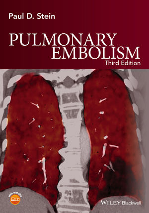
Pulmonary Embolism
Wiley-Blackwell (Verlag)
978-1-119-03908-2 (ISBN)
- Lieferbar (Termin unbekannt)
- Versandkostenfrei innerhalb Deutschlands
- Auch auf Rechnung
- Verfügbarkeit in der Filiale vor Ort prüfen
- Artikel merken
Highly illustrated with numerous tables and graphs alongside clear concise text
Includes chapters addressing pulmonary embolism (PE) and deep venous thrombosis (DVT) in relation to diseases and disorders such as; chronic heart failure, cancer, diabetes, stroke, chronic obstructive pulmonary disease (COPD) and many more
Discusses the role the different tools offered in imaging for PE, including echocardiography, multidetector computed tomography (CT), single photon emission computed tomography (SPECT), ventilation-perfusion (V-Q) imaging, dual energy CT, and magnetic resonance angiography
Contains 29 new chapters and includes new content on epidemiology of deep venous thrombosis; use of the new anticoagulants (dabigatran, rivaroxaban, and apixaban) for DVT and PE; indications and results with thrombolytic therapy and with vena cava filters; and information and indications for invasive mechanical thrombectomy and thrombolysis
Written by an internationally recognized and respected expert in the field
Paul D. Stein MD,Professor of Osteopathic Medical Specialties, College of Osteopathic Medicine, Michigan State University, East Lansing, Michigan, USA. Dr. Stein's major research in recent years has been in the field of venous thromboembolism. Dr. Stein initiated the PIOPED II and PIOPED III national collaborative studies and was national principal investigator and chairperson of the steering committees. He has written over 240 articles on venous thromboembolism from among over 560 peer reviewed articles. Dr Stein is a past president of the Laennec Society and of the American College of Chest Physicians. He is Fellow of the American College of Physicians and the American College of Cardiology and a Master Fellow of the American College of Chest Physicians. He is also a Fellow of the American Society of Mechanical Engineers. Fellowship is reserved for those who have made a significant contribution to the field of mechanical engineering. He received the Lifetime Achievement Award from the American Heart Association Midwest Affiliate, the Laureate Award of the American College of Physicians, Michigan Chapter, the Daniel Drake Award from the University of Cincinnati College of Medicine, and the Research Excellence Award from the Michigan State University College of Osteopathic Medicine. Dr. Stein also wrote a book, A Physical and Physiological Basis for the Interpretation of Cardiac Auscultation: Evaluations Based Primarily on Second Sound and Ejection Murmurs.
Prologue xi
Preface to the Third Edition xiii
About the companion website xv
Introduction 1
Part I Prevalence, risks, and prognosis of pulmonary embolism and deep venous thrombosis
1 Pulmonary embolism and deep venous thrombosis at autopsy 5
2 Incidence of pulmonary embolism and deep venous thrombosis in hospitalized patients and in emergency departments 18
3 Case fatality rate and population mortality rate from pulmonary embolism and deep venous thrombosis 24
4 Prognosis in acute pulmonary embolism based on right ventricular enlargement and biochemical markers in stable patients 31
5 Prognosis in acute pulmonary embolism based on scoring systems 43
6 Pulmonary embolism following deep venous thrombosis and outcome with untreated pulmonary embolism 49
7 Resolution of pulmonary embolism 54
8 Upper extremity deep venous thrombosis 61
9 Thromboembolic disease involving the superior vena cava and brachiocephalic veins 66
10 Venous thromboembolic disease in the four seasons 69
11 Regional differences in the United States of rates of diagnosis of pulmonary embolism and deep venous thrombosis and mortality from pulmonary embolism 73
12 Venous thromboembolism according to age and in the elderly 78
13 Pulmonary thromboembolism in infants and children 95
14 Venous thromboembolism in men and women 99
15 Pulmonary embolism and deep venous thrombosis in blacks and whites 103
16 Pulmonary thromboembolism in Asians/Pacific Islanders 108
17 Pulmonary thromboembolism in American Indians and Alaskan Natives 116
18 Venous thromboembolism in patients with cancer 118
19 Venous thromboembolism in patients with heart failure 128
20 Obesity as a risk factor in venous thromboembolism 133
21 Hypertension, smoking, and cholesterol 139
22 Overlap of venous and arterial thrombosis risk factors 141
23 Venous thromboembolism in patients with ischemic and hemorrhagic stroke 143
24 Paradoxical embolism 146
25 Pulmonary embolism and deep venous thrombosis in hospitalized adults with chronic obstructive pulmonary disease 149
26 Pulmonary embolism and deep venous thrombosis in hospitalized patients with asthma 156
27 Deep venous thrombosis and pulmonary embolism in hospitalized patients with sickle cell disease 158
28 Diabetes mellitus and risk of venous thromboembolism 162
29 Risk of venous thromboembolism with rheumatoid arthritis 164
30 Venous thromboembolism with inflammatory bowel disease 166
31 Venous thromboembolism with chronic liver disease 168
32 Nephrotic syndrome 171
33 Human immunodeficiency virus infection 173
34 Venous thromboembolism in pregnancy 176
35 Amniotic fluid embolism 182
36 Air travel as a risk for pulmonary embolism and deep venous thrombosis 184
37 Estrogen-containing oral contraceptives and venous thromboembolism 187
38 Estrogen and testosterone in men 192
39 Tamoxifen 194
40 Venous thromboembolism following bariatric surgery 198
41 Hypercoagulable syndrome 204
Part II Diagnosis of deep venous thrombosis
42 Deep venous thrombosis of the lower extremities: clinical evaluation 215
43 Clinical scoring system for assessment of deep venous thrombosis 220
44 Clinical probability score plus single negative ultrasound for exclusion of deep venous thrombosis 223
45 D-dimer for the exclusion of acute deep venous thrombosis 225
46 D-dimer combined with clinical probability assessment for exclusion of acute deep venous thrombosis 234
47 D-dimer and single negative compression ultrasound for exclusion of deep venous thrombosis 236
48 Contrast venography 237
49 Compression ultrasound for the diagnosis of deep venous thrombosis 240
50 Impedance plethysmography and fibrinogen uptake tests for diagnosis of deep venous thrombosis 247
51 Ascending CT venography and venous phase CT venography for diagnosis of deep venous thrombosis 250
52 Magnetic resonance venography for diagnosis of deep venous thrombosis 255
53 P-selectin and microparticles to predict deep venous thrombosis 260
Part III Diagnosis of acute pulmonary embolism
54 Clinical characteristics of patients with no prior cardiopulmonary disease 265
55 Relation of right-sided pressures to clinical characteristics of patients with no prior cardiopulmonary disease 272
56 The history and physical examination in all patients irrespective of prior cardiopulmonary disease 275
57 Clinical characteristics of patients with acute pulmonary embolism stratified according to their presenting syndromes 280
58 Clinical assessment in the critically ill 286
59 The electrocardiogram 289
60 The plain chest radiograph 303
61 Arterial blood gases and the alveolar–arterial oxygen difference in acute pulmonary embolism 308
62 Fever in acute pulmonary embolism 316
63 Leukocytosis in acute pulmonary embolism 319
64 Alveolar dead-space in the diagnosis of pulmonary embolism 321
65 Empirical assessment and clinical models for diagnosis of acute pulmonary embolism 324
66 Prognostic models for pulmonary embolism 329
67 D-dimer for the exclusion of acute pulmonary embolism 335
68 D-dimer combined with clinical probability for exclusion of acute pulmonary embolism 346
69 D-dimer in combination with amino-terminal pro-B-type natriuretic peptide for exclusion of acute pulmonary embolism 349
70 Tissue plasminogen activator, plasminogen activator inhibitor-1, and thrombin – antithrombin III complexes in the exclusion of acute pulmonary embolism 350
71 Echocardiogram in the diagnosis of acute pulmonary embolism 352
72 Trends in the use of diagnostic imaging in patients hospitalized with acute pulmonary embolism 356
73 Techniques of perfusion and ventilation imaging 358
74 Ventilation – perfusion lung scan criteria for interpretation prior to the Prospective Investigation of Pulmonary Embolism Diagnosis (PIOPED) 363
75 Observations from PIOPED: ventilation – perfusion lung scans alone and in combination with clinical assessment 367
76 Ventilation – perfusion lung scans according to complexity of lung disease 374
77 Perfusion lung scans alone in acute pulmonary embolism 376
78 Probability interpretation of ventilation – perfusion lung scans in relation to the largest pulmonary arterial branches in which pulmonary embolism is observed 379
79 Revised criteria for evaluation of lung scans recommended by nuclear physicians in PIOPED 381
80 Criteria for very-low-probability interpretation of ventilation – perfusion lung scans 385
81 Probability assessment based on the number of mismatched segmental equivalent perfusion defects 391
82 Probability assessment based on the number of mismatched vascular defects and stratification according to prior cardiopulmonary disease 395
83 The addition of clinical assessment to stratification according to prior cardiopulmonary disease further optimizes the interpretation of ventilation – perfusion lung scans 401
84 Pulmonary scintigraphy scans since PIOPED 407
85 Single photon emission computed tomographic (SPECT) lung scans 412
86 SPECT with radiolabeled markers 426
87 Standard and augmented techniques in pulmonary angiography 427
88 Subsegmental pulmonary embolism 435
89 Quantification of pulmonary embolism by conventional and CT angiography 440
90 Complications of pulmonary angiography 442
91 Contrast-enhanced spiral CT for the diagnosis of acute pulmonary embolism before the Prospective Investigation of Pulmonary Embolism Diagnosis II 446
92 Methods of PIOPED II 458
93 Multidetector spiral CT of the chest for acute pulmonary embolism: results of the PIOPED II trial 467
94 Multidetector CT pulmonary angiography since PIOPED II 473
95 Outcome studies of pulmonary embolism versus accuracy 478
96 Contrast-induced nephropathy 480
97 Radiation exposure and risk 483
98 Magnetic resonance angiography for the diagnosis of acute pulmonary embolism 490
99 Serial noninvasive leg tests in patients with suspected pulmonary embolism 499
100 Diagnosis of pulmonary embolism in the coronary care unit 501
101 Silent pulmonary embolism with deep venous thrombosis 506
102 Fat embolism syndrome 511
103 Diagnostic approach to acute pulmonary embolism 516
Part IV Prevention and treatment of deep venous thrombosis and pulmonary embolism
104 Warfarin and other vitamin K antagonists 523
105 Unfractionated heparin, low-molecular-weight heparin, heparinoid, and pentasaccharide 531
106 Parenteral inhibitors of factors Va, VIIIa, tissue factor, and thrombin 540
107 Novel oral anticoagulants 545
108 Aspirin for venous thromboembolism 552
109 Immediate therapeutic levels of heparin in relation to timing of recurrent events 555
110 Intermittent pneumatic compression 558
111 Graduated compression stockings 561
112 Ankle exercise and venous blood velocity 565
113 Thrombolytic therapy for deep venous thrombosis 567
114 Mechanical and ultrasonic enhancement of catheter-directed thrombolytic therapy for deep venous thrombosis 572
115 Thrombolytic therapy for treatment of acute pulmonary embolism 574
116 Catheter-tip embolectomy in the management of acute massive pulmonary embolism 589
117 Vena cava filters 597
118 Withholding treatment of patients with acute pulmonary embolism who have a high risk of bleeding provided and negative serial noninvasive leg tests 615
119 Home treatment of deep venous thrombosis 617
120 Home treatment of acute pulmonary embolism 622
121 Pulmonary embolectomy 626
122 Chronic thromboembolic pulmonary hypertension and pulmonary thromboendarterectomy 634
123 Prevention and treatment of deep venous thrombosis and acute pulmonary embolism: American College of Chest Physicians Guidelines 639
Index 647
| Erscheinungsdatum | 28.05.2016 |
|---|---|
| Verlagsort | Hoboken |
| Sprache | englisch |
| Maße | 175 x 252 mm |
| Gewicht | 1270 g |
| Themenwelt | Medizinische Fachgebiete ► Innere Medizin ► Kardiologie / Angiologie |
| Medizinische Fachgebiete ► Innere Medizin ► Pneumologie | |
| ISBN-10 | 1-119-03908-8 / 1119039088 |
| ISBN-13 | 978-1-119-03908-2 / 9781119039082 |
| Zustand | Neuware |
| Haben Sie eine Frage zum Produkt? |
aus dem Bereich


