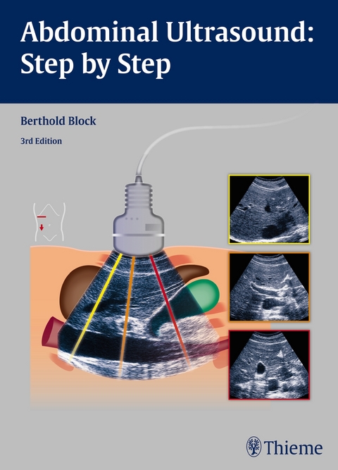
Abdominal Ultrasound: Step by Step
Seiten
2015
|
3rd edition
Thieme (Verlag)
978-3-13-138363-1 (ISBN)
Thieme (Verlag)
978-3-13-138363-1 (ISBN)
Abdominal Ultrasound: Step by Step
The third edition of this practical reference guide has been updated with a modern, visually attractive design and expanded content. The book is ideal for healthcare professionals with little or no experience in administering and interpreting abdominal ultrasound examinations. It is practice-oriented and structured in a way that allows readers with varying degrees of ultra-sonography knowledge to utilize the material according to their individual experience and needs.
Each chapter includes a systematic, detailed description of the anatomy involved in the ultrasound examination, with easy-to-digest steps that follow standardized routine and protocol. That straight-forward approach, coupled with more than 1,000 high-quality images and illustrations, enables hands-on learning, yielding the ability to assimilate these techniques quickly and adeptly.
This is a stellar resource that provides the requisite tools to locate and display the anatomical structure being tested, position and move the transducers accurately, describe and interpret the findings correctly, and differentiate key findings from the many image artifacts that typically occur.
Key Highlights:
The step-by-step guide is an invaluable, pragmatic resource to have on hand while performing abdominal ultrasound on the patient. In-depth but concise, this is an essential teaching guide for medical students, residents, technicians, and physicians who need to learn and master these examination techniques.
The third edition of this practical reference guide has been updated with a modern, visually attractive design and expanded content. The book is ideal for healthcare professionals with little or no experience in administering and interpreting abdominal ultrasound examinations. It is practice-oriented and structured in a way that allows readers with varying degrees of ultra-sonography knowledge to utilize the material according to their individual experience and needs.
Each chapter includes a systematic, detailed description of the anatomy involved in the ultrasound examination, with easy-to-digest steps that follow standardized routine and protocol. That straight-forward approach, coupled with more than 1,000 high-quality images and illustrations, enables hands-on learning, yielding the ability to assimilate these techniques quickly and adeptly.
This is a stellar resource that provides the requisite tools to locate and display the anatomical structure being tested, position and move the transducers accurately, describe and interpret the findings correctly, and differentiate key findings from the many image artifacts that typically occur.
Key Highlights:
- In-depth discussion of organ boundaries, organ details, anatomical relationships, potentially abnormal findings, tips, and clearly defined learning objectives
- Anatomical drawings incorporate a "sliced 3-D" view that show how the structures are displayed by the sector-shaper beam
- Each chapter includes a series of images replicating the 3-D impression that results from the transducer moving across the body
- Schematic drawings illustrate the ultrasound images, including a body marker that shows the transducer position
- The "sono-consultant": a systematic guide to evaluating ultrasound findings and establishing a differential diagnosis
The step-by-step guide is an invaluable, pragmatic resource to have on hand while performing abdominal ultrasound on the patient. In-depth but concise, this is an essential teaching guide for medical students, residents, technicians, and physicians who need to learn and master these examination techniques.
Berthold Block, MD, is in Private Practice, Braunschweig, Germany.
General Basics
1 General
2 Basic Physical and Technical Principles
Abdominal Ultrasound
3 Blood Vessels: The Aorta and Its Branches, the Vena Cava and Its Tributaries
4 Liver
5 Porta Hepatis
6 Gallbladder
7 Pancreas
8 Stomach, Duodenum, and Diaphragm
9 Spleen
10 Kidneys
11 Adrenal Glands
12 Bladder, Prostate, and Uterus
Brief Instructions and Documentation
13 Quick Guide
14 The Sono Consultant
15 Documentation
| Erscheinungsdatum | 17.02.2016 |
|---|---|
| Übersetzer | Terry C. Telger |
| Zusatzinfo | 1035 Abbildungen |
| Verlagsort | New York |
| Sprache | englisch |
| Maße | 195 x 270 mm |
| Gewicht | 960 g |
| Einbandart | kartoniert |
| Themenwelt | Medizinische Fachgebiete ► Radiologie / Bildgebende Verfahren ► Sonographie / Echokardiographie |
| Schlagworte | Abdomen • Bauch • Bauch / Abdomen • Bildgebendes Verfahren • Gynäkologie • gynecology • imaging procedure • Innere Medizin • Internal Medicine • Lehrbuch • Medical Studies • Medizinstudium • Pädiatrische Radiologie • Pneumologie • Radiologie • Radiologie / Nuklearmedizin • Radiology • Textbook • Ultraschall • Ultraschalldiagnostik • Ultrasonic |
| ISBN-10 | 3-13-138363-1 / 3131383631 |
| ISBN-13 | 978-3-13-138363-1 / 9783131383631 |
| Zustand | Neuware |
| Haben Sie eine Frage zum Produkt? |
Mehr entdecken
aus dem Bereich
aus dem Bereich
Begleitbuch für Sonografiekurse, Klinik und Praxis
Buch | Softcover (2023)
Urban & Fischer in Elsevier (Verlag)
27,00 €
Organbezogene Darstellung von Grund- und Aufbaukurs sowie …
Buch | Hardcover (2020)
Deutscher Ärzteverlag
99,99 €


