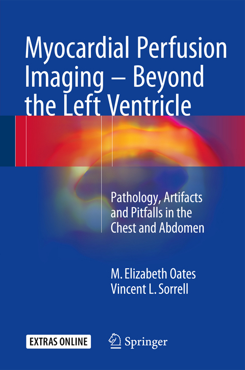
Myocardial Perfusion Imaging - Beyond the Left Ventricle
Springer International Publishing (Verlag)
978-3-319-25434-0 (ISBN)
M. Elizabeth Oates, MD, graduated from Boston University School of Medicine, Massachusetts in 1981 and returned to Boston in 1985 to complete a Nuclear Radiology fellowship at Tufts University School of Medicine/New England Medical Center (now Tufts Medical Center). During 15 years at Tufts, she rose to the rank of Professor and served as Division Chief of Nuclear Medicine. In 2001, Dr. Oates became Section Head of Nuclear Radiology at Boston Medical Center and in 2006 she accepted a position as Medical Director of Radiology at the University of Massachusetts Medical School/UMass Memorial Medical Center. In 2007, she was recruited to the University of Kentucky as Chair of the Department of Radiology where she is a tenured Professor of Radiology and Medicine (Cardiovascular Medicine) and holds the Rosenbaum Endowed Chair of Radiology. Dr. Oates is a Past President of the American Association for Women Radiologists. Honored with its Distinguished Service Award, Lifetime Service Award, and Volunteer Service Award, she currently serves on the Board of Trustees of The American Board of Radiology. She has chaired the American College of Radiology's Commission on Nuclear Medicine and Molecular Imaging since 2011 and is in her second term on the Board of Chancellors. Dr. Oates has served on both the Education Exhibits and Scientific Program Committees for the Radiological Society of North America. She is a member of the editorial board of the Journal of the American College of Radiology and recently completed service as an Associate Editor for Radiology. Dr. Oates has published 140 scholarly articles and book chapters. She has been named by Best Doctors in America annually since 2007. Dr. Oates is respected as a thought leader in her field and her opinion is regularly sought by practitioners, educators, researchers, and leaders.
Part 1 The Fundamentals: Acquisition.- Processing.- Interpretation.- Part 2 "Hot" and "Cold" Findings: Right Heart.- 1. Right atrium.- 2. Right ventricle.- Chest.- 1. Skeleton.- 2. Chest wall.- 3. Thyroid gland.- 4. Parathyroid glands.- 5. Breasts.- 6. Mediastinum.- 7. Lungs and pleura.- 8. Pericardium.- 9. Vascular system.- 10. Lymphatic system.- Abdomen.- 1. Peritoneum.- 2. Liver.- 3. Gallbladder.- 4. Spleen.- 5. Stomach.- 6. Small bowel.- 7. Large bowel.- 8. Kidneys.- 9. Vascular system.- 10. Lymphatic system.- Part 3 Case Challenges (for self-assessment)
"This book provides a review of incidental pathology, artifacts, and noncardiac findings in the chest and abdomen by myocardial perfusion imaging. ... It is a unique and important contribution to the field of myocardial perfusion imaging, and it is well done with nearly 30 well-illustrated chapters. ... Overall, this is a useful book for noninvasive cardiologists and an important contribution to the field of nuclear cardiology." (Ryan Houk, Doody's Book Reviews, February, 2017)
| Erscheinungsdatum | 13.09.2016 |
|---|---|
| Zusatzinfo | XVII, 385 p. 135 illus. in color. With online files/update. |
| Verlagsort | Cham |
| Sprache | englisch |
| Maße | 155 x 235 mm |
| Themenwelt | Medizinische Fachgebiete ► Innere Medizin ► Kardiologie / Angiologie |
| Medizinische Fachgebiete ► Radiologie / Bildgebende Verfahren ► Nuklearmedizin | |
| Medizinische Fachgebiete ► Radiologie / Bildgebende Verfahren ► Radiologie | |
| Medizin / Pharmazie ► Studium | |
| Schlagworte | Cardiology • diagnostic radiology • heart • Medicine • Myokardperfusion • Nuclear Medicine • Radionuclide imaging • SPECT • Stress Test • Technetium-99m • Thallium-201 |
| ISBN-10 | 3-319-25434-0 / 3319254340 |
| ISBN-13 | 978-3-319-25434-0 / 9783319254340 |
| Zustand | Neuware |
| Haben Sie eine Frage zum Produkt? |
aus dem Bereich


