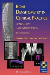
Bone Densitometry in Clinical Practice
Humana Press Inc. (Verlag)
978-0-89603-513-3 (ISBN)
- Titel erscheint in neuer Auflage
- Artikel merken
This unique summary of the state-of-the-art in X-ray bone densitometry highlights for today's primary care physicians and densitometrists the expert application of densitometry techniques in patient management. The book describes the various densitometry techniques currently in use and how to intelligently select among them in a wide variety of clinical circumstances. It also shows how to interpret the computer-generated data and how to recognize and avoid interpretation pitfalls from the effects of artifacts and structural changes. Physicians performing densitometry will learn the key elements to include in a densitometry report to referring doctors, and how to gain mastery of the sophisticated quality control procedures necessary for the successful performance of bone densitometry. Offering the most comprehensive but easily readable explanation of diagnostic densitometry for the physician and technologist, Bone Densitometry in Clinical Practice will become the essential guide for all medical personnel working with densitometry.
Chapter 1. Densitometry Techniques in Medicine Today. Plain Radiography in the Assessment of Bone Density. Qualitative Spinal Morphometry and the Singh Index. Qualitative Spinal Morphometry. The Singh Index. Quantitative Morphometric Techniques: Calcar Femorale Thickness Radiogrammetry and the Radiologic Osteoporosis Score . Calcar Femorale Thickness. Radiogrammetry. The Radiologic Osteoporosis Score. Radiographic Photodensitometry. Radiographic Absorptiometry (RA). Photon Absorptiometric Techniques. Single-Photon Absorptiometry (SPA). Dual-Photon Absorptiometry (DPA). Dual-Energy X-Ray Absorptiometry (DXA). Peripheral DXA Units. DTX-200. pDEXA. Peripheral Instantaneous X-Ray Imager (PIXI). Single-Energy X-Ray Absorptiometry (SXA). Quantitative Computed Tomography (QCT). Peripheral QCT. References. Chapter 2: Densitometric Anatomy. The Skeleton in Densitometry. Axial and Appendicular Skeleton.Weight-Bearing and Non-Weight Bearing Skeleton. Central and Peripheral Skeleton. Skeletal Site Composition. The Spine in Densitometry. Vertebral Anatomy. Artifacts in AP Spine Densitometry. Vertebral Fractures. Effect of Osteophytes on BMD. Effect of Aortic Calcification on BMD. Effect of Facet Sclerosis on BMD. Other Causes of Artifacts in AP Spine Studies. The Spine in the Lateral Projection. The Proximal Femur in Densitometry. Proximal Femur Anatomy. Effect of Rotation on BMD in the Proximal Femur. Effect of Leg Dominance on BMD in the Proximal Femur. Effect of Artifacts on BMD in the Proximal Femur. The Forearm in Densitometry . Nomenclature. Effect of Arm Dominance on Forearm BMD. Effect of Artifacts on BMD in the Forearm. Other Skeletal Sites. References. Chapter 3. Statistics in Densitometry. Mean Variance and Standard Deviation. The Mean. Variance and Standard Deviation. Coefficient of Variation. Standard Scores. Z-Scores. T-Scores. Measures of Risk. Prevalence and Incidence. Prevalence. Incidence. Absolute Relative and Attributable Risk. Absolute Risk. Relative Risk. Attributable Risk. Odds Ratios. Confidence Intervals. Accuracy and Precision. Accuracy. Precision. Correlation. Statistical Significance and the P-Value. References. Chapter 4. The Importance of Precision in Densitometry. Performing a Short-Term Precision Study. Mathematical Procedures Used to Calculate Precision. Applying the Precision Value to the Interpretation of Serial Measurements. The Confidence Interval for the Change in BMD Between Two Measurements. Effect of Precision on the Timing of Repeat Measurements of BMD. References. Chapter 5. Quality-Control Procedures for Densitometry. Establishing a Baseline Value with the Phantom. Shewhart Rules and CUSUM Charts. Shewhart Rules. CUSUM Charts. Automated Quality Control Procedures. References. Chapter 6. The Prediction of Fracture Risk with Densitometry. Prevalence of Fracture at Different Levels of BMD. Fracture-Risk Prediction. Site-Specific and Global Fracture Risk Prediction. Relative-Risk Fracture Data. Global Fracture-Risk Prediction. Site-Specific Spine Fracture-Risk Prediction. Site-Specific Hip Fracture-Risk Prediction. Applying Relative-Risk Data in Clinical Practice. Lifetime Risk of Fracture. Remaining- Lifetime Fracture Probability. Fracture Threshold. Other Risk Assessments Derived from or Combined with Densitometry. Pre-Existing Fractures. Increasing Number of Low Bone-Mass Sites. Hip-Axis Length. References. Chapter 7. The Effects of Age Disease and Drugs on Bone Density. Age-Related Changes in Bone Density. Bone Density in Children. Bone Density in Premenopausal Women. Dissimilar BMDs Between Skeletal Sites at Peak and Prior to Menopause. Bone Density in Perimenopausal Women. Dissimilar Spine and Femoral BMD in Perimenopausal Women. Changes in Bone Density in Postmenopausal Women. Changes in Bone Density in Men. Diseases Known to Affect Bone Density. Acromegaly. Alcoholism. Amenorrhea. Hyperandrogenic Amenorrhea Exercise- Induced Amenorrhea. Anorexia Nervosa. Cirrhosis. Diabetes. Insulin-Dependent Diabetes Mellitus. Noninsulin-Dependent Diabetes Mellitus (NIDDM). Estrogen Deficiency (Postmenopausal). Gastrectomy . Gaucher Disease Type I. Glutlen-Sensitive Enteropathy. Human Immunodeficiency Virus Infection. Hypercalciuria. Hyperparathyroidism. Hyperprolactinemia. Hyperthyroidism. Inflammatory Bowel Disease. Intravenous Drug Abuse. Klinefelter's Syndrome. Marfan's Syndrome. Mastocytosis. Multiple Myeloma. Multiple Sclerosis.Osteoarthritis. Paralysis. Hemiplegia. Paraplegia. Parkinson's Disease. Pregnancy. Renal Failure. Rheumatoid Arthritis. Thalassemia Major. Transient Osteoporosis of the Hip. Transplantation. Cardiac Transplantation. Marrow Transplantation. Renal Transplantation. Effect of Drugs on Bone Density. Alendronate Sodium. Calcitriol. Calcium and Vitamin D. Corticosteroids. Oral Corticosteroids. Inhaled Corticosteroids. Estrogen/Hormone Replacement. Etidronate. GnRH Agonists. Heparin. Ipriflavone. Medroxyprogesterone Acetate. Nandrolone Decanoate. Raloxifene. Risedronate. Salmon Calcitonin. Sodium Fluoride. Tamoxifen. Thyroid Hormone. Tibolone. References. Chapter 8. Bone Density Data from DPA to DXA and Manufacturer to Manufacturer. From DPA to DXA. Hologic DXA and Lunar DPA. Lunar DXA and Lunar DPA. DXA: From Lunar to Hologic to Norland. Hologic DXA and Norland DXA. Lunar DXA and Hologic DXA. Standardization of Absolute BMD Results. From DXA Machine to DXA Machine Within Manufacturers. From Pencil-Beam to Fan-Array DXA Data. Reference Databases. References. Chapter 9. Clinical Indications for Bone Densitometry. Clinical Guidelines. 1988 National Osteoporosis Foundation Guidelines. Guidelines of the International Society for Clinical Densitometry. 1996 American Association of Clinical Endocrinologists' Guidelines. Guidelines from the European Foundation for Osteoporosis and Bone Disease. Guidelines of the Study Group of the World Health Organization for the Diagosis of Osteoporosis. How Do the Guidelines Compare? Clinical Applications of Bone Mass Measurements. Selection of Sites to Measure. Assessment of Fracture Risk. Quantification of Bone Density. Confirmation of Suspected Demineralization or Skeletal Fragility. Diagnosis of Osteoporosis. Assessment of the Effect of Disease Processes on the Skeleton. Serial Assessment of Bone Density to Assess the Effects of Disease or Therapeutic Interventions. General Considerations in Site Selection. References. Chapter 10. Case Studies in Densitometry. Index.
| Erscheint lt. Verlag | 24.6.1998 |
|---|---|
| Reihe/Serie | Current Clinical Practice |
| Verlagsort | Totowa, NJ |
| Sprache | englisch |
| Maße | 229 x 152 mm |
| Gewicht | 698 g |
| Themenwelt | Medizinische Fachgebiete ► Chirurgie ► Unfallchirurgie / Orthopädie |
| Medizinische Fachgebiete ► Innere Medizin ► Endokrinologie | |
| ISBN-10 | 0-89603-513-1 / 0896035131 |
| ISBN-13 | 978-0-89603-513-3 / 9780896035133 |
| Zustand | Neuware |
| Haben Sie eine Frage zum Produkt? |
aus dem Bereich


