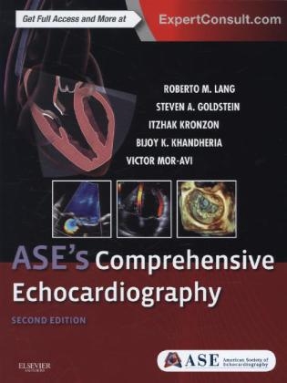
ASE's Comprehensive Echocardiography
Elsevier - Health Sciences Division (Verlag)
978-0-323-26011-4 (ISBN)
- Titel erscheint in neuer Auflage
- Artikel merken
"I am also very proud that this textbook illustrates what is great about the ASE. We are a society with more than 16,000 members worldwide, dedicated to quality in cardiovascular ultrasound and education, both of which are prominently demonstrated throughout this textbook. ASE is also a village made up of many different people from many different backgrounds, all united and energized about the value of cardiovascular ultrasound in caring for people worldwide." Foreword by: Neil J. Weissman, American Society of Echocardiography, July 2015
Take advantage of an outstanding online library of slides and videos of case presentations that correspond to crisp, full-color images, allowing you to view dynamic echocardiographic clips of various cardiac pathologies.
Tap into the knowledge and skills of a team of experts from the ASE, led by world-renowned authorities in echocardiography.
Access the fully searchable text online at expertconsult.com, along with additional cases, images, and an extensive library of cine images.
Get fully up to date with the latest echo practice guidelines and advanced technologies, including 3D echocardiography and myocardial strain.
Gain a better understanding of the latest methods to assess cardiac chamber size and function, valvular stenosis/regurgitation, cardiomyopathies, coronary artery disease, complications of myocardial infarction, and much more - all in a practical, well-illustrated brief yet comprehensive format extensively supported by multimedia material.
Stay up to date with hot topics in this rapidly evolving field: interventional/intraoperative echocardiography, transesophageal echocardiography, cardiac resynchronization therapy, and more.
Set the pace with enhanced technologies and guidelines
Section I. Physics and Instrumentation
1. General Principles of Echocardiography
2. Three-Dimensional Echocardiography
3. Doppler Principles
4. Tissue Doppler Imaging and Speckle Tracking Echocardiography
5. Tissue Harmonic Imaging
Section II. Transthoracic Echocardiography
6. Transthoracic Echocardiography: Nomenclature and standard views
7. Technical quality
8. Transthoracic Echocardiography Tomographic Views
9. M- Mode Echocardiography
10. Doppler Echocardiography: Normal Antegrade Flow Patterns
Section III. Transesophageal Echocardiography
11. Protocol, Probe Insertion and Manipulation, Risks and Complications
12. Transesophageal Echocardiography: Tomographic View
13. Applications of Transesophageal Echocardiography
14. Pitfalls and Artifacts in Transesophageal Echocardiography
Section IV. Intracardiac Echocardiography
15. Applications of Intracardiac Echocardiography
16. Limitations of Intracardiac Echocardiography
Section V. Intravascular Echocardiography
17. Intravascular Ultrasound: Instrumentation and Technique
18. Intravascular Ultrasound: Applications and Limitations
Section VI. Hand-Held Echocardiography
19. Hand-carried cardiac ultrasound: Background, Instrumentation and Technique
20. Focused Cardiac Ultrasound
Section VII. Contrast Echocardiography
21. Contrast Echocardiography: Introduction
22. Ultrasonic contrast agents
23. Physical Properties of Microbubble Ultrasound Contrast Agents
24. Applications of Ultrasound Contrast Agents
25. Stress Echocardiography and Contrast
26. Contrast-enhanced carotid imaging
Section VIII. Left Ventricular Systolic Function
27. Introduction
28. Left Ventricular Systolic function: Basic Principles
29. Global Left Ventricular systolic function
30. Regional Left Ventricular Systolic Function
31. Assessment of left ventricular dyssynchrony
Section IX. Right Heart
32. Right Ventricular Anatomy
33. The Physiologic Basis of Right Ventricular Echocardiography
34. Assessment of Right Ventricular Systolic and Diastolic Function
35. Right Ventricular Hemodynamics
36. The right atrium
37. Pulmonary embolism
Section X. Diastolic Function
38. Physiology of diastole
39. Methods of Assessment
40. Echo Doppler parameters of diastolic function
41. Estimation of Left Ventricular filling pressures
42. Clinical Recommendations for Echocardiography Laboratories for Assessment of Left Ventricular Diastolic Function
43. Newer Methods to Assess Diastolic Function
44. Causes of diastolic dysfunction
Section XI. Left Atrium
45. Assessment of Left Atrial Size
46. Assessment of Left Atrial Function
Section XII. Ischemic Heart Disease
47. Introduction to Ischemic Heart Disease
48. Ischemic Heart Disease: Basic Principles
49. Acute Chest Pain Syndromes: Differential Diagnosis
50. Echocardiography in Acute Myocardial Infarction
51. Echocardiography in Stable Coronary Artery Disease
52. Old Myocardial Infarction
53. End-Stage Cardiomyopathy Due to Coronary Artery Disease
54. Coronary artery anomalies
Section XIII. Stress Echocardiography
55. Stress Echocardiography - Introduction
56. Effects of Exercise, Pharmacological Stress and Pacing on the Cardiovascular System
57. Diagnostic criteria, accuracy
58. Stress Echocardiography Methodology
59. Stress Echocardiography: Image acquisition
60. Prognosis
61. Viability
62. Contrast-enhanced stress echocardiography
63. 3Dimensional stress echocardiography
64. Stress Echocardiography for Valve disease: Aortic Regurgitation and Mitral Stenosis
65. Appropriate Use Criteria for Stress Echocardiography
66. Comparison with other techniques
Section XIV. Cardiomyopathies
67. Introduction to Cardiomyopathies
68. Pathophysiology and Variants of Hypertrophic Cardiomyopathy
69. Hypertrophic Cardiomyopathy: Pathophysiology, Functional Features and Treatment of Outflow Tract Obstruction
70. Differential of Hypertrophic Cardiomyopathy versus Secondary Conditions that Mimic Hypertrophic Cardiomyopathy
71. Echocardiographic Features of Hypertrophic Cardiomyopathy: Mechanism of Systolic Anterior Motion
72. Hypertrophic cardiomyopathy: Assessment of therapy
73. Hypertrophic cardiomyopathy: Screening of relatives
74. Apical hypertrophic cardiomyopathy
75. Echocardiography in Athletic Preparticipation Screening
76. Dilated Cardiomyopathy: Etiology, Diagnostic Criteria and Echocardiographic Features
77. Imaging in Familial Dilated Cardiomyopathy
78. Echocardiographic Predictors of Outcome in Patients with Dilated Cardiomyopathy
79. Right Ventricle in Dilated Cardiomyopathy
80. Restrictive cardiomyopathy: Classification
81. Cardiac Amyloidosis - Echocardiographic Features
82. Hereditary and Acquired Infiltrative Cardiomyopathy
83. Endomyocardial Fibrosis
84. Restriction versus constriction
85. Echocardiography in Arrhythmogenic Right Ventricular Cardiomyopathy
86. Echocardiographic Analysis of Left Ventricular Noncompaction
87. Takotsubo-like Transient Left Ventricular Dysfunction: Takotsubo Cardiomyopathy
88. A Systematic Echocardiographic Approach to Left Ventricular Assist Device Therapy
89. Posttransplantation echocardiographic evaluation
90. Familial Cardiomyopathies
91. Echocardiography in Cor Pulmonale and/or Pulmonary heart disease
92. Echocardiographic Evaluation of Functional Tricuspid Regurgitation
93. Echocardiographic Evaluation of the Right Heart: Limitations and Technical Considerations
Section XV. Aortic Stenosis
94. Aortic stenosis morphology
95. Quantification of aortic stenosis severity
96. Asymptomatic severe aortic stenosis
97. Risk stratification - timing of surgery
98. Low-Flow, low gradient, Aortic Stenosis with Reduced Left Ventricular Ejection Fraction
99. Low-Flow, low gradient, Aortic Stenosis with Preserved Left Ventricular Ejection Fraction
100. Stress (Exercise) Echocardiography in Asymptomatic Aortic Stenosis
101. Subaortic Stenosis
Section XVI. Aortic Regurgitation
102. Introduction to Aortic Regurgitation
103. Aortic Regurgitation: Etiologies and Left Ventricular Responses
104. Aortic Regurgitation: Pathophysiology
105. Quantitation of Aortic Regurgitation
106. Risk stratification - timing of surgery for aortic regurgitation
Section XVII. Mitral Stenosis
107. Mitral Stenosis: Introduction
108. Rheumatic mitral stenosis
109. Quantification of Mitral Stenosis
110. Other (nonrheumatic) etiologies of Mitral Stenosis; situations that mimic Mitral Stenosis
111. Role of hemodynamic stress testing in Mitral Stenosis
112. Consequences of Mitral Stenosis
Section XVIII. Mitral Regurgitation
113. Introduction to Mitral Regurgitation
114. Etiologies and Mechanisms of Mitral Valve Dysfunction
115. Mitral valve prolapse
116. Quantification of Mitral Regurgitation
117. Asymptomatic severe Mitral Regurgitation
118. Role of exercise stress testing
119. Ischemic Mitral Regurgitation
Section XIX. Tricuspid Regurgitation
120. Epidemiology, Etiology and Natural History of Tricuspid Regurgitation
121. Quantification of Tricuspid Regurgitation
122. Indications for Tricuspid Valve Surgery
123. Tricuspid Valve Procedures
Section XX. Pulmonic Regurgitation
124. Introduction and Etiology of Pulmonic Regurgitation
125. Pulmonic Regurgitation: Semiquantification
Section XXI. Prosthetic Valves
126. Prosthetic Valves: Introduction
127. Classification of Prosthetic valve types and fluid dynamics
128. Aortic prosthetic valves
129. Mitral prosthetic valves
130. Periprosthetic leaks
131. Tricuspid prosthetic valves
132. Mitral valve repair
Section XXII. Infective Endocarditis
133. Introduction and Echocardiographic Features of Infective Endocarditis
134. Infective Endocarditis: Role of Transthoracic versus Transesophageal Echocardiography
135. Echocardiography for Prediction of Cardioembolic Risk
136. Echocardiography and Decision Making for Surgery
137. Intraoperative Echocardiography in Infective Endocarditis
138. Limitations and Technical considerations
Section XXIII. Pericardial Diseases
139. Introduction to Pericardial diseases
140. Normal pericardial anatomy
141. Pericarditis
142. Pericardial effusion and cardiac tamponade
143. Constrictive pericarditis
144. Effusive Constrictive pericarditis
145. Pericardial cysts and Congenital absence of pericardium
Section XXIV. Tumors and Masses
146. Introduction to Echocardiographic Assessment of Cardiac Tumors and Masses
147. Primary benign, malignant, and metastatic tumors in the heart
148. Left Ventricular thrombus
149. Left Atrial thrombus
150. Right heart thrombus
151. Normal anatomic variants and artifacts
152. Role of contrast echocardiography in the assessment of intracardiac masses
153. Echocardiography-guided biopsy of intracardiac masses
154. Cardiac sources of emboli
Section XXV. Diseases of the Aorta
155. Introduction
156. Aortic atherosclerosis and Embolic Events
157. Aortic aneurysm
158. Sinus of Valsalva aneurysm
159. Acute aortic syndrome
160. Penetrating Atherosclerotic Ulcer and Intramural hematoma
161. Aortic trauma
162. Intraoperative echocardiography
163. Postoperative echocardiography of the Aorta
Section XXVI. Adult Congenital Heart Disease
164. Introduction
165. Systematic approach to adult Congenital Heart Disease
166. Common Congenital Heart Defects Associated with left-to-Right Shunts
167. Obstructive Lesions
168. The Adult with Unrepaired Complex Congenital Heart Defects
169. Adult Congenital heart disease with prior surgical repair
Section XXVII. Systemic Diseases
170. Hypertension
171. Diabetes
172. End-stage renal disease
173. Obesity
174. Rheumatic fever and Rheumatic Heart Disease
175. Systemic Lupus Erythematosus
176. Antiphospholipid antibody syndrome
177. Carcinoid heart Disease
178. Amyloid
179. Sarcoidosis
180. Cardiac Involvement in Hypereosinophilic Syndrome
181. Endocrine disease
182. Chagas Cardiomyopathy
183. Sickle cell disease
184. Human Immunodeficiency Virus
185. Cardiotoxic effects of cancer therapy
186. Pregnacy and the heart
187. Cocaine
Section XXVIII. Echocardiography in the Emergency Room
188. Echocardiography in Emergency clinical presentation
Section XXIX. Interventional Echocardiography
189. Introduction
190. Transcatheter aortic valve replacement
191. MitraClip Procedure
192. Mitral balloon valvuloplasty
193. Transcatheter valve in valve implantation
194. Atrial and Ventricular Septal Defect Closure
195. Transcatheter Cardiac pseudoaneurysm closure
196. Patent foramen ovale
197. Fusion of Three-Dimensional echocardiography with fluoroscopy for intervention guidance
Section XXX. Miscellaneous Topics in Echocardiography
198. Appropriate use criteria
199. Carotid Ultrasound to Evaulate Cardiovascular Disease Risk: Carotid Intima-Media Thickness and Plaque Detection
200. Coronary artery imaging
| Erscheint lt. Verlag | 19.5.2015 |
|---|---|
| Zusatzinfo | Approx. 950 illustrations (600 in full color); Illustrations |
| Verlagsort | Philadelphia |
| Sprache | englisch |
| Gewicht | 2790 g |
| Themenwelt | Medizinische Fachgebiete ► Innere Medizin ► Kardiologie / Angiologie |
| Medizinische Fachgebiete ► Radiologie / Bildgebende Verfahren ► Sonographie / Echokardiographie | |
| ISBN-10 | 0-323-26011-X / 032326011X |
| ISBN-13 | 978-0-323-26011-4 / 9780323260114 |
| Zustand | Neuware |
| Haben Sie eine Frage zum Produkt? |
aus dem Bereich


