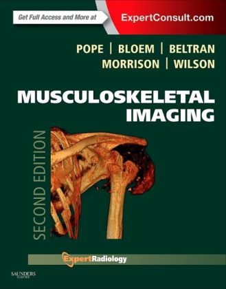
Musculoskeletal Imaging
Saunders (Verlag)
978-1-4557-0813-0 (ISBN)
- Titel ist leider vergriffen;
keine Neuauflage - Artikel merken
"This is an excellent benchbook and accompanying electronic resource which will be of value to trainee radiologists and established consultants."� Reviewed by: Dr Steve Amerasekara, Consultant Radiologist on behalf of journal RAD Magazine, July 2015
"This outstanding text is now an acclaimed primary resource and therefore belongs in the libraries and at the work stations of all general and orthopedic hospital departments of radiology and, indeed, at any and all imaging facilities involved in musculoskeletal imaging." Foreword�by: Lee F. Rogers,��June 2015
Fully understand each topic with a format that delivers essential background information.
Streamline the decision-making process with integrated protocols, classic signs, and ACR guidelines, as well as a design that structures every chapter consistently to include pathophysiology, imaging techniques, imaging findings, differential diagnosis, and treatment options.
Write the most comprehensive reports possible with help from boxes highlighting what the referring physician needs to know, as well as suggestions for treatment and future imaging studies.
Access in-depth case studies, valuable appendices, and additional chapters covering all of the most important musculoskeletal procedures performed today.
Quickly locate important information with a full-color design that includes color-coded tables and bulleted lists highlighting key concepts, as well as color artwork that lets you easily find critical anatomic views of diseases and injuries.
Engage with more than 40 brand-new videos, including arthroscopic videos.
Easily comprehend complicated material with over 5,000 images and new animations.
Explore integrated clinical perspectives on the newest modalities such as PET-CT in cancer, diffusion MR, as well as ultrasonography, fusion imaging, multi-slice CT and nuclear medicine.
Learn from team of international experts provides a variety of evidence-based guidance, including the pros and cons of each modality, to help you overcome difficult challenges.
Expert Consult eBook version included with purchase. This enhanced eBook experience allows you to search all of the text, figures, references, and videos from the book on a variety of devices.
1. General Imaging Principles
PART I: INJURYSection 1: Axial Skeleton 2. General Principles of Osseous Injury 3. Imaging of Facial and Skull Trauma 4. Cervical Spine Injuries 5. Injury of the Thoracic Cage and Thoracolumbar Spine Section 2: Appendicular Skeleton Upper Extremities 6. Normal Shoulder 7. Osseous Injuries of the Shoulder Girdle 8. Shoulder Impingement Syndromes 9. Glenohumeral Instability 10. Normal Elbow 11. Acute Osseous Injury of the Elbow and Forearm 12. Soft Tissue Injury to the Elbow 13. Normal Wrist 14. Acute Osseous Injury to the Wrist 15. Internal Derangement of the Wrist 16. Acute Osseous�Trauma to�the Hand 17. Compressive and Entrapment Neuropathies of the Upper Extremities 18. Soft Tissue Injuries of the Hand and Wrist
Lower Extremities 19. Normal Pelvis and Hip 20. Acute Osseous Injury�to the Pelvis and Acetabulum 21. Athletic Pubalgia 22. Acute Osseous Injury to the Hip and Proximal Femur 23. Internal Derangement of the Hip and Proximal Femur 24. Normal Knee 25. Acute Osseous Injury to the Knee 26. Internal Derangement of the Knee: Meniscal�Injuries 27. Internal Derangement of the Knee:�Ligament Injuries 28. Internal Derangement of the Knee: Tendon Injuries 29. Internal Derangement of the Knee: Cartilage and Osteochondral Injuries 30. Normal Ankle and Foot 31. Acute Osseous Injury to the Ankle and Foot 32. Soft Tissue Injury to the Ankle: Ligament Injuries 33. Soft Tissue Injury to the Ankle: Tendon Injuries 34. Soft Tissue Injury to the Ankle: Osteochondral Injuries and Impingement 35.�Compressive and Entrapment Neuropathies�of the Lower Extremity 36. Imaging of the Forefoot
Section 3: Pediatric Injuries 37. Lower Extremity Injuries in Children 38. Upper Extremity Injuries in Children 39. Skeletal Manifestations of Pediatric Nonaccidental Injury Section 4: Other Musculoskeletal Injuries 40. Stress Injury 41. Radiation Effects in the Musculoskeletal System 42. Complications of Osseous Trauma 43. Muscle Injury and Sequelae 44. Complex Regional Pain Syndrome PART II: ARTHROPATHIES AND NEUROLOGIC/MUSCULAR DISORDERS AND CONNECTIVE TISSUE DISEASE 45. Degenerative Disorders of the Spine 46. Aging 47. Degenerative Disease: Physiology and Advanced Imaging 48. Rheumatoid Arthritis 49. Psoriatic Arthritis and Psoriatic Spondyloarthropathy 50. Reactive Arthritis 51. Ankylosing Spondylitis 52. Progressive Scleroderma 53. Systemic Lupus Erythematosus 54. Mixed Connective Tissue Disease 55. Juvenile Idiopathic Arthritis 56. Idiopathic Inflammatory Myopathy 57. Hemochromatosis 58. Ochronosis 59. Diffuse Idiopathic Skeletal Hyperostosis and Ossification of the Posterior Longitudinal Ligament 60. Gout 61. Crystal-Related Arthritis 62. Neuropathic Osteoarthropathy PART III: INFECTION 63. Noninflammatory Intraarticular Pathology 64. Soft Tissue Infection: Cellulitis, Pyomyositis, Abscess, Septic Arthritis 65. Appendicular Infection 66. Spinal Infection 67. Diabetic Pedal Infection 68. Pediatric Infections 69. HIV Infection and AIDS 70. Atypical Mycobacterial Infection PART IV: HEMATOLOGIC AND VASCULAR DISEASE 71. General Principles of MRI of the Bone Marrow 72. Ischemic Bone Lesions 73. Hemophilia and Related Disorders 74. Sickle Cell Anemia 75. Thalassemia 76. Myelofibrosis PART V: METABOLIC, HORMONAL, AND SYSTEMIC DISEASE 77. Osteoporosis 78. Hyperparathyroidism, Renal Osteodystrophy, Osteomalacia, and Rickets 79. Amyloidosis 80. Pituitary and Thyroid Disorders 81. Gaucher Disease 82. Storage Diseases 83. Osteogenesis Imperfecta 84. Marfan Syndrome 85. Paget Disease 86. Hypertrophic Osteoarthropathy 87. Sarcoidosis 88. Tuberous Sclerosis 89. Drug-Related Bone and Soft Tissue Disorders PART VI: MUSCULOSKELETAL TUMORS AND TUMOR-LIKE LESIONS 90. The Patient with a Tumor or a Tumor-Like Lesion of Bone 91. The Patient with a Soft Tissue Lump 92. Primary Bone Tumors 93. Myeloma 94. Tumor-Like Lesions of Bone 95. Soft Tissue Tumors 96. Tumor-Like Soft Tissue Lesions 97. Metastatic Disease 98. Treatment Strategies for Musculoskeletal Tumors and Tumor-Like Lesions 99. Staging Bone and Soft Tissue Tumors 100. Monitoring Therapy in Bone and Soft Tissue Tumors PART VII: CLINICALLY RELEVANT DEVELOPMENT DYSPLASIAS 101. Focal Growth Disturbances 102. Developmental Dysplasia of the Hip 103. Coalitions 104. Dysplasias 105. Spinal Deformity PART VIII: POSTSURGICAL IMAGING AND COMPLICATIONS 106. Principles and Complications of Orthopedic Hardware 107. Postoperative Shoulder 108. Postoperative Elbow, Wrist, and Hand 109. Postoperative Hip 110. Postoperative Knee 111. Postoperative Ankle and Foot 112. Imaging of the Residual Limb after Amputation 113. Postoperative Infections PART IX: MISCELLANEOUS 114. Temporomandibular Joint 115. Dental Imaging 116. Normal Variants
PART IX: MUSCULOSKELETAL PROCEDURES 117. Biopsy: Soft Tissue 118. Percutaneous Biopsy of the Appendicular Skeleton 119. Percutaneous Biopsy of the Spine 120. Tumor Ablation 121. Spinal Injections 122. Discography 123. Vertebroplasty and Kyphoplasty 124. Percutaneous Intradiskal Therapies 125. Ultrasound Procedures APPENDICES Appendix 1: Measurements Most Frequently Used in Orthopedic Imaging Appendix 2: Orthopedic Devices Appendix 3: Fractures with Names Appencix 4: Diseases with Names Appendix 5: Classic Signs�and Findings in�Musculoskeletal Radiology
| Reihe/Serie | Expert Radiology |
|---|---|
| Zusatzinfo | Approx. 6000 illustrations |
| Verlagsort | Philadelphia |
| Sprache | englisch |
| Maße | 222 x 281 mm |
| Themenwelt | Medizin / Pharmazie ► Medizinische Fachgebiete ► Orthopädie |
| Medizinische Fachgebiete ► Radiologie / Bildgebende Verfahren ► Nuklearmedizin | |
| ISBN-10 | 1-4557-0813-5 / 1455708135 |
| ISBN-13 | 978-1-4557-0813-0 / 9781455708130 |
| Zustand | Neuware |
| Haben Sie eine Frage zum Produkt? |
aus dem Bereich


