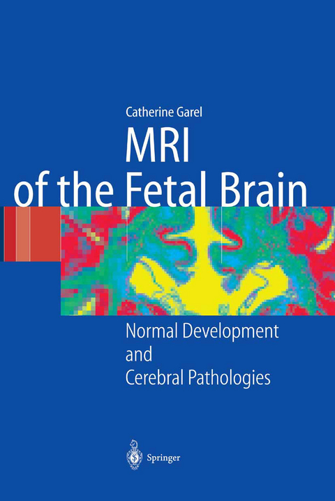
MRI of the Fetal Brain
Springer Berlin (Verlag)
978-3-642-62275-5 (ISBN)
Fetal MR has grown continually in importance in recent years, and the brain has become the main focus of investigation. However, we lack established standards and a good knowledge of the normal MR appearance. To fill this gap is the purpose of the first part of this book, which is an MR atlas of the cerebral development of the fetus.
The second part is dedicated to cerebral pathologies. It includes, for each condition, a summary of the fundamental data, the imaging findings (US and MR) in correlation with neurofetopathology and/or postnatal imaging, and a brief perspective of the prognosis.
1 Techniques.- 2 Materials.- 3 Methodology.- 4 Results.- 5 Comments on the Results.- 6 Appendix.- 7 References.- 8 Pathology of the Midline.- 9 Abnormalities of Proliferation, Neuronal Migration and Cortical Organization.- 10 Intracranial Space-Occupying Lesions.- 11 Ventricular Dilatation.- 12 Abnormalities of the Posterior Cerebral Fossa.- 13 Antenatal Cerebral Pathologies of Infectious Origin.- 14 Abnormalities of the Fetal Cerebral Parenchyma: Ischaemic and Haemorrhagic Lesions.- 263.
From the reviews:
"An extremely organised and well presented tome. ... forms two parts. The first part is an atlas of the development of the fetal brain. The sequences are correlated with pathological specimens and are clearly labelled and are very impressive. The second part is, I think, more relevant to clinical radiologists ... . images really are excellent and, again, detail is clear and the description of the pathology is excellent. ... it will guide you through protocols ... . In all, an excellent book ... ." (Dr. N S Ashford, RAD Magazine, August, 2005)
"The editor, Catherine Garel, presents a book on the development of the fetal brain and cerebral pathologies using magnetic resonance imaging (MRI). ... The book is characterized by its many images, its clear structure and the good description of the findings. ... In summary, the present book offers insight into the development of fetal MRI, focusing on the brain. It can be recommended to both practitioners and professionals." (T. J. Vogl, European Radiology, Vol. 16, 2006)
| Erscheint lt. Verlag | 22.11.2012 |
|---|---|
| Co-Autor | A.-L. Delezoide, L. Guibaud, G. Sebag, P. Gressens, M. Elmaleh-Bergès, M. Hassan, H. Brisse, E. Chantrel |
| Übersetzer | V. Delezoide |
| Zusatzinfo | XV, 267 p. |
| Verlagsort | Berlin |
| Sprache | englisch |
| Original-Titel | 1. Garel, C. - Imagerie du cerveau foetal pathologique 2. Garel, C. - Developpement du cerveau foetal |
| Maße | 193 x 270 mm |
| Gewicht | 620 g |
| Themenwelt | Medizin / Pharmazie ► Gesundheitsfachberufe ► Hebamme / Entbindungspfleger |
| Medizin / Pharmazie ► Medizinische Fachgebiete ► Pädiatrie | |
| Medizinische Fachgebiete ► Radiologie / Bildgebende Verfahren ► Radiologie | |
| Schlagworte | brain • Brain: Growth and development • Fetal abnormalities • Fetal brain • Fetal central nervous system • Fetal MR • Fetus • growth • Magnetic Resonance Imaging (MRI) |
| ISBN-10 | 3-642-62275-5 / 3642622755 |
| ISBN-13 | 978-3-642-62275-5 / 9783642622755 |
| Zustand | Neuware |
| Haben Sie eine Frage zum Produkt? |
aus dem Bereich


