Atlas of Fundus Fluorescein Angiography
Jaypee Brothers Medical Publishers (Verlag)
978-93-5025-577-3 (ISBN)
- Titel z.Zt. nicht lieferbar
- Versandkostenfrei innerhalb Deutschlands
- Auch auf Rechnung
- Verfügbarkeit in der Filiale vor Ort prüfen
- Artikel merken
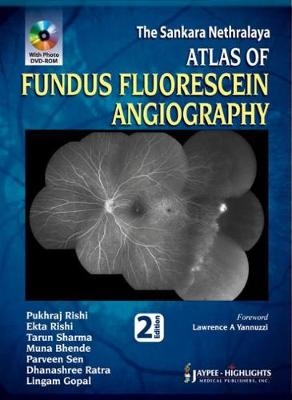
Fundus fluorescein angiography is a technique for examining the circulation of the retina using a dye tracing method.
This atlas is a comprehensive guide to fluorescein angiographic findings of ocular disorders. Beginning with an introduction to the technique, the following sections discuss macular disorders, vascular disorders, inflammatory disorders, heredomacular dystrophy, optic nerve disorders and tumours.
Featuring more than 1400 colour images, this second edition includes a free photo CD of case studies, as well as a self test chapter for quick revision.
Key points
Comprehensive guide to fundus fluorescein angiography
Includes free photo CD of case studies
More than 1400 colour images
Self test chapter for quick revision
Previous edition published 2009
Pukhraj Rishi MS DO Ekta Rishi MS Tarun Sharma MD FRCSEd MBA Muna Bhende MS Parveen Sen MS Dhanashree Ratra MS DNB FRCSEd Lingam Gopal MS DNB FRCSEd All at Sri Bhagwan Mahavir, Vitreoretinal Service, Sankara Nethralaya, Chennai, Tamil Nadu, India
Section 1: Getting Started
Introduction • Checklist for Fluorescein Angiography • Photographic and Angiographic Artifacts • Fundus Autofluorescence • Normal Fluorescein Angiogram • Abnormal Fluorescence
Section 2: Macular Disorders
Drusen • Epiretinal Membrane • Macular Hole • Myopia • Pigment Epithelial Detachment • Age-related Macular Degeneration—Dry Type • Age-related Macular Degeneration—Wet Type • Retinal Angiomatous Proliferans • Polypoidal Choroidal Vasculopathy • Choroidal Neovascular Membranes (Other than ARMD) • Retinal Pigment Epithelial Rip • Choroidal Rupture • Angioid Streaks • Choroidal Folds • Acute Macular Neuroretinopathy • Central Serous Chorioretinopathy • Drug Toxicity • Photic Maculopathy • Cystoid Macular Edema
Section 3: Vascular Disorders
Arterial Occlusion • Coats’ Disease • Diabetic Retinopathy • Venous Occlusion • Ocular Ischemic Syndrome • Eales’ Disease • Familial Exudative Vitreoretinopathy • Hypertensive Retinopathy • Macroaneurysm • Parafoveal Telangiectasia • Purtscher’s Retinopathy • Valsalva Retinopathy •
Radiation Retinopathy
Section 4: Inflammatory Disorders
Acute Retinal Pigment Epitheliitis (Krill’s disease) • Acute Posterior Multifocal Placoid Pigment • Epitheliopathy • Geographic Helicoid Peripapillary Choroidopathy • Vogt-Koyanagi-Harada Syndrome • Sympathetic Ophthalmitis • Behçet’s Syndrome • Intermediate Uveitis • Birdshot Retinochoroidopathy • Multiple Evanescent White Dot Syndrome • Punctate Inner Choroidopathy • Acute Retinal Necrosis • Posterior Scleritis • Choroidal Granuloma • Other Inflammatory Disorders
Section 5: Heredomacular Dystrophy
Bietti’s Crystalline Dystrophy • Best’s Disease • Stargardt’s Disease (Fundus Flavimaculatus) • Progressive Cone Dystrophy • Other Heredomacular Dystrophies • Ocular Albinism • Retinitis Pigmentosa and Allied Disorders
Section 6: Optic Nerve Disorders
Optic Nerve Disorders
Section 7: Tumors
Choroidal Nevus • Melanocytoma • Malignant Melanoma • Choroidal Hemangioma • Choroidal Osteoma • Retinoblastoma • Retinal Angiomatosis • Astrocytic Hamartoma • Other Tumors • Self Test
Glossary
Index
| Erscheint lt. Verlag | 31.3.2013 |
|---|---|
| Zusatzinfo | 1422 Halftones, color |
| Verlagsort | New Delhi |
| Sprache | englisch |
| Maße | 216 x 279 mm |
| Gewicht | 1500 g |
| Themenwelt | Medizin / Pharmazie ► Medizinische Fachgebiete ► Augenheilkunde |
| Medizin / Pharmazie ► Medizinische Fachgebiete ► Radiologie / Bildgebende Verfahren | |
| ISBN-10 | 93-5025-577-4 / 9350255774 |
| ISBN-13 | 978-93-5025-577-3 / 9789350255773 |
| Zustand | Neuware |
| Haben Sie eine Frage zum Produkt? |
aus dem Bereich


