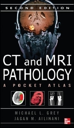
CT & MRI Pathology: A Pocket Atlas, Second Edition
Seiten
2012
|
2nd edition
McGraw-Hill Medical (Verlag)
978-0-07-170319-2 (ISBN)
McGraw-Hill Medical (Verlag)
978-0-07-170319-2 (ISBN)
- Titel ist leider vergriffen;
keine Neuauflage - Artikel merken
Zu diesem Artikel existiert eine Nachauflage
Reviews the pathology, etiology, signs and symptoms, imaging characteristics, treatment, and prognosis for each disease/disorder and includes crisp images to accompany every discussion. This title features: written by a radiographic technologist and a radiologist; images are organized by body system for easy reference; and detailed summaries.
A pocket atlas of 180 of the most common pathologies visualized on CT and MRI
CT & MRI Pathology, 2e is a portable reference of 180 common pathologies seen in CT and on MRI. It concisely reviews the pathology, etiology, signs and symptoms, imaging characteristics, treatment, and prognosis for each disease/disorder and includes crisp, high-quality images to accompany every discussion.
New to the second edition are an additional 90 disease topics with new images.
Features
Written by a radiographic technologist and a radiologistImages are organized by body system for easy referenceDetailed summaries
Michael L.Grey, MS RT(R)(MR)(CT) is certified in MR and CT and teaches in the radiologic technology program at Southern Illinois University in Carbondale.
Part I: Principles of Imaging in Pathology; Part II: Central Nervous System; Part III: Head and Neck; Part IV: Chest and Mediastinum; Part V: Abdomen; Part VI: Pelvis; Part VII: Musculoskeletal; Bibliography; Index
| Zusatzinfo | 400 Illustrations |
|---|---|
| Verlagsort | New York |
| Sprache | englisch |
| Maße | 119 x 216 mm |
| Gewicht | 455 g |
| Themenwelt | Medizinische Fachgebiete ► Radiologie / Bildgebende Verfahren ► Computertomographie |
| Medizinische Fachgebiete ► Radiologie / Bildgebende Verfahren ► Kernspintomographie (MRT) | |
| Studium ► 2. Studienabschnitt (Klinik) ► Pathologie | |
| ISBN-10 | 0-07-170319-5 / 0071703195 |
| ISBN-13 | 978-0-07-170319-2 / 9780071703192 |
| Zustand | Neuware |
| Haben Sie eine Frage zum Produkt? |
Mehr entdecken
aus dem Bereich
aus dem Bereich


