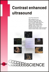
Contrast enhanced ultrasound
UNI-MED (Verlag)
978-3-8374-1221-5 (ISBN)
- Titel erscheint in neuer Auflage
- Artikel merken
This book was written by internationally renowned experts in this field and provides up-to-date information about the principles of CEUS and the indications and interpretation in different organ diseases when using US contrast agents.
1.Basics13
1.1.Bubbles and how ultrasound systems talk to them (P.N. Burns)13
1.1.1.Contrast agents for ultrasound14
1.1.1.1.Blood pool agents14
1.1.1.2.Selective uptake agents15
1.1.2.Talking to bubbles: physical principles of contrast imaging16
1.1.2.1.Bubble behavior and incident pressure16
1.1.2.2.The Mechanical Index (MI)16
1.1.2.3.Nonlinear echoes and harmonic imaging17
1.1.2.4.Harmonic B-mode imaging18
1.1.2.5.Pulse inversion imaging18
1.1.2.6.Pulse inversion Doppler imaging: amplitude and phase modulation19
1.1.2.7.Temporal Maximum Intensity Projection (MIP) imaging20
1.1.2.8.Disrupting bubbles: intermittent imaging20
1.1.3.References21
1.2.Safety considerations and regulatory status (P.N. Burns)23
1.2.1.References25
1.3.Clinical aspects of severe adverse reactions (M. Höpfner)26
1.3.1.References26
1.4.CEUS image documentation (L. Thorelius)27
1.4.1.CEUS and the time factor27
1.4.2.Documentation basics27
1.4.3.Characterization of FLL’s27
1.4.4.Detection of liver metastases28
1.4.5.Non-liver CEUS documentation31
1.5.Examination strategies, tips & techniques (H.-P. Weskott)31
1.5.1.Prior to a CEUS study31
1.5.2.Reasons for an insufficient contrast enhancement33
1.5.3.Dosage of contrast agent35
1.5.4.Acoustic output (MI) and bubble destruction36
1.5.5.Probe types and contrast imaging37
1.5.6.CEUS performance39
1.5.7.References43
2.Liver44
2.1.Kinetics of US contrast agents (H.-P. Weskott)44
2.2.Benign focal liver lesions (K.F. Stock)45
2.2.1.Benign cystic liver lesions46
2.2.2.Focal fatty infiltration46
2.2.3.Hemangioma47
2.2.4.Focal nodular hyperplasia48
2.2.5.Hepatic adenoma50
2.2.6.Rare benign focal liver lesions52
2.2.7.References52
2.3.Infectious diseases (L. Thorelius)54
2.3.1.Liver infections54
2.3.2.Liver abscesses54
2.3.3.Renal infections56
2.3.4.Infections elsewhere57
2.3.5.Endocavitary CEUS58
2.4.Hepatocellular carcinoma (R. Lencioni, C. Della Pina, L. Crocetti)58
2.4.1.Introduction58
2.4.2.Ultrasound and Contrast-enhanced Ultrasound59
2.4.3.Diagnostic work-up62
2.4.4.References63
2.5.Cholangiocarcinoma (C. Pachmann)65
2.5.1.References67
2.6.Detection of focal liver lesions (H.-P. Weskott)68
2.6.1.References71
2.7.Differentiation between benign and malignant liver lesions (H.-P. Weskott)72
2.7.1.References77
2.8.Evaluation and characterization of metastatic liver disease (H.-P. Weskott)77
2.8.1.Vascular involvement82
2.8.2.References86
2.9.The role of contrast enhanced ultrasound in liver surgery (E. Leen, G. Low)86
2.9.1.Introduction86
2.9.2.Background87
2.9.3.Contrast enhanced intraoperative US (CE-IOUS) technique88
2.9.4.Characterization of focal liver lesions with CE-IOUS89
2.9.5.CE-IOUS of liver metastases90
2.9.6.Actual change in surgical management as a result of CE-IOUS91
2.9.7.Clinical perspective91
2.9.8.CE-IOUS of HCC92
2.9.9.Limitations of CE-IOUS94
2.9.10.Future directions and challenges94
2.9.11.References94
2.10.Evaluation of anti-angiogenic treatments using dynamic contrast-enhanced ultrasonography with quantification (N. Lassau, L. Chami, M. Chebil)97
2.10.1.Post processing98
2.10.2.Further evaluation of DCE-US98
2.10.3.References105
3.Gallbladder diseases (H.-P. Weskott)107
3.1.Cholecystitis and complications107
3.2.Malignant tumors of the gallbladder wall111
3.3.References112
4.Pancreas114
4.1.Pancreatic vasculature and parenchymal enhancement (M. D’Onofrio, A. Gallotti, R.P. Mucelli)114
4.2.Comparison to other imaging modalities (M. D’Onofrio, A. Gallotti, R.P. Mucelli)114
4.3.Examination technique (M. D’Onofrio, A. Gallotti, R.P. Mucelli)116
4.4.Inflammatory diseases (M. D’Onofrio, A. Gallotti, R.P. Mucelli)116
4.5.Cystic tumors (M. D’Onofrio, A. Gallotti, R.P. Mucelli)117
4.6.Solid pancreatic tumors (C. Pachmann)119
4.7.References122
5.Spleen (C. Görg)125
5.1.Splenic vasculature and parenchymal enhancement125
5.2.Examination technique125
5.3.Diffuse splenic diseases126
5.4.Vascular splenic lesions129
5.5.Benign focal lesions131
5.6.Secundary malignant focal splenic lesions134
5.7.References135
6.Kidneys and collective system137
6.1.Infectious diseases (H.-P. Weskott)137
6.2.Solid renal tumors (H.-P. Weskott)141
6.2.1.Benign renal tumors143
6.2.1.1.Angiomyolipoma143
6.2.1.2.Oncocytoma146
6.2.2.Malignant renal tumors148
6.3.Cystic renal tumors (H.-P. Weskott)153
6.3.1.Urothelium carcinoma156
6.3.2.Renal lymphomas158
6.3.3.References (Chapter 6.1.-6.3.)159
6.4.Disorders of the renal blood supply (K.F. Stock, H.-P. Weskott)161
6.4.1.References165
6.5.Transplant kidney (K.F. Stock)166
6.5.1.References169
6.6.Monitoring of tumor treatment: CEUS in the management of thermal ablation in renal cell cancer (D. Clevert)170
6.6.1.Introduction170
6.6.2.Pre-Interventional, peri-interventional and follow-up imaging and the potential of Contrast-Enhanced Ultrasound (CEUS)171
6.6.3.References175
7.Abdominal aortic pathologies and follow up after endovascular aneurysm repair (EVAR) (D. Clevert)177
7.1.Normal abdominal aortic anatomy177
7.2.Abdominal aortic aneurysm (AAA)177
7.3.Aortic dissections178
7.4.Aorto-caval fistulas180
7.5.Inflammatory abdominal aortic aneurysm180
7.6.Endovascular aneurysm repair (EVAR)182
7.7.References185
8.Neovascularization of the vessel wall: Role and diagnostic approach toneovascularization in vascular disease (H.-P. Weskott)188
8.1.Examination technique188
8.2.CEUS in arterial lumen imaging189
8.3.Vessel wall and the process of vascularization190
8.4.References194
Index
| Reihe/Serie | UNI-MED Science |
|---|---|
| Zusatzinfo | 230 illustrations |
| Sprache | englisch |
| Maße | 170 x 240 mm |
| Gewicht | 510 g |
| Einbandart | gebunden |
| Themenwelt | Medizin / Pharmazie ► Medizinische Fachgebiete |
| Schlagworte | Ultraschalldiagnostik |
| ISBN-10 | 3-8374-1221-0 / 3837412210 |
| ISBN-13 | 978-3-8374-1221-5 / 9783837412215 |
| Zustand | Neuware |
| Haben Sie eine Frage zum Produkt? |
aus dem Bereich