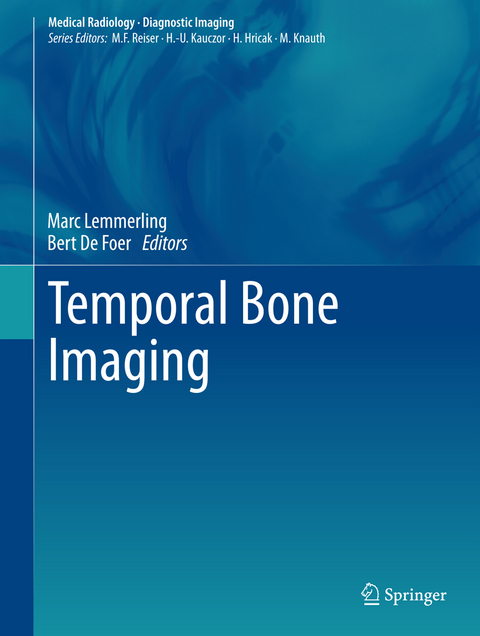
Temporal Bone Imaging
Seiten
2014
|
2015
Springer Berlin (Verlag)
978-3-642-17895-5 (ISBN)
Springer Berlin (Verlag)
978-3-642-17895-5 (ISBN)
This thorough overview of imaging of normal and diseased temporal bone covers both classic methods and newer techniques such as functional imaging and diffusion-weighted imaging. Includes diseases and conditions likely to be encountered in clinical practice.
This book provides a complete overview of imaging of normal and diseased temporal bone. After description of indications for imaging and the cross-sectional imaging anatomy of the area, subsequent chapters address the various diseases and conditions that affect the temporal bone and are likely to be encountered regularly in clinical practice. The classic imaging methods are described and discussed in detail, and individual chapters are included on newer techniques such as functional imaging and diffusion-weighted imaging. There is also a strong focus on postoperative imaging. Throughout, imaging findings are documented with the aid of numerous informative, high-quality illustrations. Temporal Bone Imaging, with its straightforward structure based essentially on topography, will prove of immense value in daily practice.
This book provides a complete overview of imaging of normal and diseased temporal bone. After description of indications for imaging and the cross-sectional imaging anatomy of the area, subsequent chapters address the various diseases and conditions that affect the temporal bone and are likely to be encountered regularly in clinical practice. The classic imaging methods are described and discussed in detail, and individual chapters are included on newer techniques such as functional imaging and diffusion-weighted imaging. There is also a strong focus on postoperative imaging. Throughout, imaging findings are documented with the aid of numerous informative, high-quality illustrations. Temporal Bone Imaging, with its straightforward structure based essentially on topography, will prove of immense value in daily practice.
Indications for temporal bone imaging.- temporal bone imaging techniques.- cross sectional imaging anatomy of the temporal bone.-external ear imaging.- acute otomastoiditis and its complications.-chronic otomastoiditis without cholesteatoma.- chronic otomastoiditis with cholesteatoma.- temporal bone trauma.- temporal bone tumours.- congenital malformations of the temporal bone.- pathology of the cerebellopontine angle and internal auditory canal.- inner ear pathology.- imaging of cochlear implants.- petrous bone apex lesions.- pathology of the facial nerve.- imaging of the jugular foramen.- vascular temporal bone lesions.- imaging of the postoperative temporal bone.- mpr ct imaging of the temporal bone.- functional imaging of hearing.
| Erscheint lt. Verlag | 17.11.2014 |
|---|---|
| Reihe/Serie | Diagnostic Imaging | Medical Radiology |
| Zusatzinfo | X, 380 p. 445 illus., 79 illus. in color. |
| Verlagsort | Berlin |
| Sprache | englisch |
| Maße | 210 x 279 mm |
| Gewicht | 1303 g |
| Themenwelt | Medizin / Pharmazie ► Medizinische Fachgebiete ► Chirurgie |
| Medizin / Pharmazie ► Medizinische Fachgebiete ► HNO-Heilkunde | |
| Medizinische Fachgebiete ► Radiologie / Bildgebende Verfahren ► Radiologie | |
| Schlagworte | Bildgebende Verfahren (Medizin) • CT • Ear Imaging • Head and Neck Surgery • Imaging • Knochen • MRI • otorhinolaryngology • otosclerosis • temporal bone |
| ISBN-10 | 3-642-17895-2 / 3642178952 |
| ISBN-13 | 978-3-642-17895-5 / 9783642178955 |
| Zustand | Neuware |
| Haben Sie eine Frage zum Produkt? |
Mehr entdecken
aus dem Bereich
aus dem Bereich
Buch (2023)
Thieme (Verlag)
190,00 €


