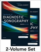
Textbook of Diagnostic Sonography
Mosby (Verlag)
978-0-323-07301-1 (ISBN)
- Titel erscheint in neuer Auflage
- Artikel merken
Stay up to date with the rapidly changing field of medical sonography! Heavily illustrated and extensively updated to reflect the latest developments in the field, Textbook of Diagnostic Sonography, 7th Edition equips you with an in-depth understanding of general/abdominal and obstetric/gynecologic sonography, the two primary divisions of sonography, as well as vascular sonography and echocardiography. Each chapter includes patient history, normal anatomy (including cross-sectional anatomy), ultrasound techniques, pathology, and related laboratory findings, giving you comprehensive insight drawn from the most current, complete information available.
Full-color presentation enhances your learning experience with vibrantly detailed images.
Pathology tables give you quick access to clinical findings, laboratory findings, sonography findings, and differential considerations.
Sonographic Findings highlight key clinical information.
Key terms and chapter objectives help you study more efficiently.
Review questions on a companion Evolve website reinforce your understanding of essential concepts.
New chapters detail the latest clinically relevant content in the areas of:
Essentials of Patient Care for the Sonographer
Artifacts in Image Acquisition
Understanding Other Imaging Modalities
Ergonomics and Musculoskeletal Issues in Sonography
3D and 4D Evaluation of Fetal Anomalies
More than 700 new images (350 in color) clarify complex anatomic concepts.
Extensive content updates reflect important changes in urinary, liver, musculoskeletal, breast, cerebrovascular, gynecological, and obstetric sonography.
Volume 1
�
Part I: FOUNDATIONS OF SONOGRAPHY
1. Foundations of Sonography
2. Introduction to Physical Findings, Physiology, and Laboratory Data
3. Essentials of Patient Care for the Sonographer NEW
4. Ergonomics and Musculoskeletal Issues in Sonography NEW
5. Understanding Other Imaging Modalities NEW
6. Artifacts in Scanning NEW
�
Part II: ABDOMEN
7. Anatomic and Physiologic Relationships within the Abdominal Cavity
8. Introduction to Abdominal Scanning: Techniques and Protocols
9. The Vascular System
10. The Liver
11. The Gallbladder and the Biliary System
12. The Pancreas
13. The Gastrointestinal Tract
14. The Urinary System
15. The Spleen
16. The Retroperitoneum
17. The Peritoneal Cavity and Abdominal Wall
18. Abdominal Applications of Ultrasound Contrast
19. Ultrasound-Guided Interventional Techniques
20. Emergent Abdominal Ultrasound Procedures
�
Part III: SUPERFICIAL STRUCTURES
21. The Breast
22. The Thyroid
23. The Scrotum
24. The Musculoskeletal System
�
Part IV: NEONATAL AND PEDIATRICS
25. Neonatal Echoencephalography
26. The Pediatric Abdomen: Jaundice and Common Surgical Conditions
27. The Neonatal and Pediatric Kidneys and Adrenal Glands
28. The Neonatal and Pediatric Pelvis
29. The Neonatal Hip
30. The Neonatal Spine
Volume 2
Part V: THE THORACIC CAVITY
31. Anatomic and Physiologic Relationships within the Thoracic Cavity
32. Introduction to Echocardiographic Evaluation and Techniques
33. Fetal Echocardiography: Beyond the Four Chambers
34. Fetal Echocardiography: Congenital Heart Disease
�
Part VI: CEREBROVASCULAR
35. Extracranial Cerebrovascular Evaluation
36. Intracranial Cerebrovascular Evaluation
37. Peripheral Arterial Evaluation
38. Peripheral Venous Evaluation
�
Part VII: GYNECOLOGY
39. Normal Anatomy and Physiology of the Female Pelvis
40. Sonographic and Doppler Evaluation of the Female Pelvis
41. Uterine Pathology
42. Ovarian Pathology
43. Adnexal Pathology
44. The Role of Sonography in Evaluating Female Infertility
�
Part VIII: OBSTETRICS
45. The Role of Sonography in Obstetrics
46. Clinical Ethics for Obstetric Sonography
47. The Normal First Trimester
48. First Trimester Complications
49. Sonography of the Second and Third Trimesters
50. Obstetric Measurements and Gestational Age
51. Fetal Growth Assessment using Sonography
52. Sonography and High Risk Pregnancy
53. Prenatal Diagnosis of Congenital Anomalies
54. 3D and 4D Evaluation of Fetal Anomalies NEW
55. The Placenta
56. The Umbilical Cord
57. Amniotic Fluid, Membranes, and Fetal Hydrops
58. The Fetal Face and Neck
59. The Fetal Neural Axis
60. The Fetal Thorax
61. The Fetal Anterior Abdominal Wall
62. The Fetal Abdomen
63. The Fetal Urogenital System
64. The Fetal Skeleton
| Zusatzinfo | Approx. 3463 illustrations (1098 in full color) |
|---|---|
| Verlagsort | St Louis |
| Sprache | englisch |
| Maße | 222 x 281 mm |
| Themenwelt | Medizinische Fachgebiete ► Radiologie / Bildgebende Verfahren ► Sonographie / Echokardiographie |
| ISBN-10 | 0-323-07301-8 / 0323073018 |
| ISBN-13 | 978-0-323-07301-1 / 9780323073011 |
| Zustand | Neuware |
| Haben Sie eine Frage zum Produkt? |
aus dem Bereich


