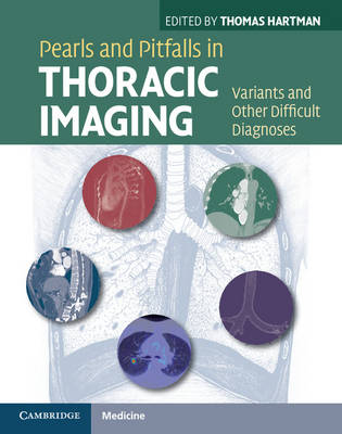
Pearls and Pitfalls in Thoracic Imaging
Variants and Other Difficult Diagnoses
Seiten
2011
Cambridge University Press (Verlag)
978-0-521-11907-8 (ISBN)
Cambridge University Press (Verlag)
978-0-521-11907-8 (ISBN)
A key diagnostic challenge for radiologists is distinguishing between normal variants (anatomic difference not indicating disease) and pathologic findings that do indicate disease. Pearls and Pitfalls in Thoracic Imaging is an invaluable resource - hundreds of images and concise descriptions enable rapid, accurate diagnosis of the full range of thoracic disorders.
How often have you been confronted with an image on a thoracic CT exam where you knew it didn't look 'normal', but you weren't sure whether it was 'abnormal' either? And if it is abnormal, is there a specific diagnosis you should be able to make directly off the images? Pearls and Pitfalls in Thoracic Imaging is your one-stop resource to answer questions such as: Is this a normal variant or a disease-related abnormality? Are these findings specific for an uncommon disease and if so what is the diagnosis? Is this set of findings strongly suggestive of a diagnosis? Which additional imaging test will allow me to be confident in that diagnosis? Could the 'abnormality' be due to an artifact mimicking disease? Written by leading thoracic radiologists and with concise, image-rich descriptions, Pearls and Pitfalls in Thoracic Imaging is an invaluable diagnostic tool for every radiologist.
How often have you been confronted with an image on a thoracic CT exam where you knew it didn't look 'normal', but you weren't sure whether it was 'abnormal' either? And if it is abnormal, is there a specific diagnosis you should be able to make directly off the images? Pearls and Pitfalls in Thoracic Imaging is your one-stop resource to answer questions such as: Is this a normal variant or a disease-related abnormality? Are these findings specific for an uncommon disease and if so what is the diagnosis? Is this set of findings strongly suggestive of a diagnosis? Which additional imaging test will allow me to be confident in that diagnosis? Could the 'abnormality' be due to an artifact mimicking disease? Written by leading thoracic radiologists and with concise, image-rich descriptions, Pearls and Pitfalls in Thoracic Imaging is an invaluable diagnostic tool for every radiologist.
Thomas Hartman is Professor of Radiology and Associate Chair for Education, Mayo Clinic Department of Radiology, Rochester, MN, USA.
Preface; Airways Thomas Hartman; Lung parenchyma David Levin; Mediastinum John Hildebrandt; Esophagus John Barlow; Aorta Patrick Eiken; Vascular Anne-Marie Sykes; Pericardium Rebecca Lindell; Pleura Rebecca Lindell; Diaphragm Rebecca Lindell; Lymphatics John Hildebrandt; PET and PET/CT Patrick Peller; Artifacts Anne-Marie Sykes; Index.
| Zusatzinfo | 12 Halftones, color; 389 Halftones, black and white |
|---|---|
| Verlagsort | Cambridge |
| Sprache | englisch |
| Maße | 223 x 283 mm |
| Gewicht | 1080 g |
| Themenwelt | Medizinische Fachgebiete ► Innere Medizin ► Pneumologie |
| Medizinische Fachgebiete ► Radiologie / Bildgebende Verfahren ► Radiologie | |
| ISBN-10 | 0-521-11907-3 / 0521119073 |
| ISBN-13 | 978-0-521-11907-8 / 9780521119078 |
| Zustand | Neuware |
| Haben Sie eine Frage zum Produkt? |
Mehr entdecken
aus dem Bereich
aus dem Bereich
Aus der Praxis für die Praxis
Buch (2022)
Thieme (Verlag)
71,00 €
International Trauma Life Support (ITLS)
Buch | Softcover (2024)
Hogrefe AG (Verlag)
70,00 €


