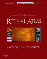
The Retinal Atlas
Expert Consult - Online and Print
Seiten
2010
W B Saunders Co Ltd (Verlag)
978-0-7020-3320-9 (ISBN)
W B Saunders Co Ltd (Verlag)
978-0-7020-3320-9 (ISBN)
- Titel erscheint in neuer Auflage
- Artikel merken
Zu diesem Artikel existiert eine Nachauflage
Offers over 5,000 illustrations of the advanced imaging and research findings essential for effective diagnosis of retinal disorders. This title is suitable for retinal specialists, comprehensive ophthalmologists, and other eye care personnel.
*2011 BMA Awards - Highly Commended in� Internal Medicine *
Dr. Lawrence A. Yannuzzi brings together the most complete retinal atlas ever. Over 5,000 illustrations of the latest imaging and research findings essential for effective diagnosis of retinal disorders populate The Retinal Atlas. A unique page layout consisting of optimally positioned panoramic images, magnified photos, and histopathological specimens illustrate key manifestations, giving you the best visual display of each disease. In addition, composite images using different retinal imaging modalities, including the latest in optical coherence tomography (OCT), fluorescein angiography, indocyanine green (ICG), and fundus autofluorescence display how a disease appears in each imaging modality, allowing you to compare imaging methods and gain a better understanding of each disorder. The Atlas is the ideal resource for all retinal specialists, comprehensive ophthalmologists, and other eye care personnel. The Expert Consult functionality gives you easy access to the full text online, as well as a downloadable image library at expertconsult.com.
Chapter 1: Normal
Chapter 2: Hereditary chorioretinal dystrophies
Chapter 3: Pediatric retina
Chapter 4: Inflammation
Chapter 5: Infection
Chapter 6: Retinal vascular disease
Chapter 7: Degeneration
Chapter 8: Oncology
Chapter 9: Macular fibrosis, pucker, cysts, holes, folds, and edema
Chapter 10: Non-rhegmatogenous retinal detachment
Chapter 11: Peripheral retinal degenerations and rhegmatogenous retinal detachment
Chapter 12: Traumatic chorioretinopathy
Chapter 13: Complications of ocular surgery
Chapter 14: Chorioretinal toxicities
Chapter 15: Congenital anomalies of the optic nerve
| Erscheint lt. Verlag | 1.6.2010 |
|---|---|
| Zusatzinfo | Approx. 5125 illustrations (3744 in full color) |
| Verlagsort | London |
| Sprache | englisch |
| Themenwelt | Medizin / Pharmazie ► Medizinische Fachgebiete ► Augenheilkunde |
| ISBN-10 | 0-7020-3320-0 / 0702033200 |
| ISBN-13 | 978-0-7020-3320-9 / 9780702033209 |
| Zustand | Neuware |
| Informationen gemäß Produktsicherheitsverordnung (GPSR) | |
| Haben Sie eine Frage zum Produkt? |
Mehr entdecken
aus dem Bereich
aus dem Bereich
Ein systematischer Ansatz
Buch | Hardcover (2022)
Urban & Fischer in Elsevier (Verlag)
265,00 €
Buch | Spiralbindung (2022)
Thieme (Verlag)
71,00 €


