Introductory Biomedical Imaging
CRC Press (Verlag)
978-1-138-62668-3 (ISBN)
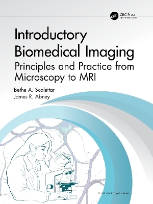
Imaging is everywhere. We use our eyes to see and cameras to take pictures. Scientists use microscopes and telescopes to peer into cells and out to space. Doctors use ultrasound, X-rays, radioisotopes, and MRI to look inside our bodies. If you are curious about imaging, open this textbook to learn the fundamentals.
Imaging is a powerful tool in fundamental and applied scientific research and also plays a crucial role in medical diagnostics, treatment, and research. This undergraduate textbook introduces cutting-edge imaging techniques and the physics underlying them. Elementary concepts from electromagnetism, optics, and modern physics are used to explain prominent forms of light microscopy, as well as endoscopy, ultrasound, projection radiography and computed tomography, radionuclide imaging, and magnetic resonance imaging. This textbook also covers digital image processing and analysis. Theoretical principles are reinforced with illustrative homework problems, applications, activities, and experiments, and by emphasizing recurring themes, including the effects of resolution, contrast, and noise on image quality. Readers will learn imaging fundamentals, diagnostic capabilities, and strengths and weaknesses of techniques.
This textbook had its genesis, and has been vetted, in a "Biomedical Imaging" course at Lewis & Clark College in Portland, OR, and is designed to facilitate the teaching of similar courses at other institutions. It is unique in its coverage of both optical microscopy and medical imaging at an intermediate level, and exceptional in its coverage of material at several levels of sophistication.
Bethe A. Scalettar is a professor and chair of physics at Lewis & Clark College in Portland, OR. She joined the College in 1993 after receiving an undergraduate degree from the University of California at Irvine (majors, physics and mathematics), a PhD in biophysics from the University of California at Berkeley, and completing postdoctoral work at the University of California, San Francisco. Since arriving at Lewis & Clark College, Bethe’s research and publications have focused primarily on elucidating the molecular basis of learning and memory, utilizing fluorescence microscopy as a primary tool. Bethe also has worked to enhance the interdisciplinary appeal of physics, most notably with the development of an undergraduate physics course entitled “Biomedical Imaging” and the writing of this supporting textbook. James R. Abney is an intellectual property attorney at Psi Star Intellectual Property LLC in Portland, OR. He began practicing law in 1997 after receiving undergraduate degrees in physics and biology from the University of California at Irvine, a PhD in biophysics from the University of California at Berkeley, completing postdoctoral work at the University of California, San Francisco, and earning a J.D. from Lewis & Clark College School of Law. Jim’s research and publications, which have continued during his legal career, have focused on the use of biophysical, notably fluorescence-based, techniques to answer questions about cell structure and function. Jim’s legal work has spanned multiple technologies useful in science and medicine, including scientific instrumentation (especially light sources, detectors, and optics), medical devices, and biotechnology.
1. Introduction, 2. Review of Essential Basics, Section I. Microscopy, 3. Introduction to Image Formation by the Optical Microscope, 4. Wave Theory of Image Formation and Resolution, 5. Contrast Enhancement in Optical Microscopy, 6. Fluorescence Microscopy, 7. Axially Selective Fluorescence Excitation Techniques, 8. Super-Resolution Fluorescence Techniques, 9. Detectors, Sampling, and Image Processing and Analysis, Section II. Medical Imaging, 10. Ultrasound, 11. Projection Radiography and Computed Tomography, 12. Planar Scintigraphy and Emission Tomography, 13. Magnetic Resonance Imaging
| Erscheinungsdatum | 18.04.2024 |
|---|---|
| Reihe/Serie | Imaging in Medical Diagnosis and Therapy |
| Zusatzinfo | 18 Tables, color; 40 Tables, black and white; 262 Line drawings, color; 4 Line drawings, black and white; 64 Halftones, color; 21 Halftones, black and white; 326 Illustrations, color; 25 Illustrations, black and white |
| Verlagsort | London |
| Sprache | englisch |
| Maße | 210 x 280 mm |
| Gewicht | 453 g |
| Themenwelt | Medizin / Pharmazie ► Physiotherapie / Ergotherapie ► Orthopädie |
| Naturwissenschaften ► Physik / Astronomie ► Angewandte Physik | |
| Technik ► Medizintechnik | |
| ISBN-10 | 1-138-62668-6 / 1138626686 |
| ISBN-13 | 978-1-138-62668-3 / 9781138626683 |
| Zustand | Neuware |
| Haben Sie eine Frage zum Produkt? |
aus dem Bereich


