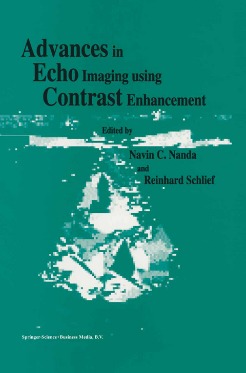
Advances in Echo Imaging Using Contrast Enhancement
Springer (Verlag)
978-94-015-8128-8 (ISBN)
When these drugs are available, sophisticated computed methodologies have to be included in the echocardio- graphic machines in order to improve the determination of the left ventricular volume and ejection fraction [44]. In the future, cineventriculography will be rarely performed as echoventriculograms already show left ventricular contraction. This will possibly result in reduced side effects and costs. REFERENCES 1. Gramiak R, Shah PM, Kramer DH. Ultrasound cardiography: Contrast studies in anatomy and function. Radiology 1969; 939. 2. Kronik G, Hutterer B, Mosslacher H. Diagnose atrialer Links-rechts-Shunts mit Hilfe der zweidimensionalen Kontrastechokardiographie. Z Kardiol 1981;70:138-45.
One: History, basics and safety of contrast agents.- 1. Contrast-echocardiography — a historical perspective.- 2. Principles of echo contrast.- 3. Conventional echo-contrast agents. Hand preparation, sonication, properties.- 4. Albumin spheres as contrast agents.- 5. Saccharide based contrast agents.- 6. Echocontrast enhancers — how safe are they?.- 7. Gas bubble dynamics in acoustic fields and their biological consequences.- Two: Clinical uses of contrast agents.- 8. Clinical uses of contrast agents — practical considerations.- 9. Structure identification by transthoracic contrast echocardiography.- 10. Identification of right sided structures by contrast transesophageal echocardiography.- 11. Left ventricular contrast echocardiography — echoventriculography.- 12. Diagnosis of patent foramen ovale by transesophageal and transthoracic echocardiography.- 13. Spontaneous echographic contrast — etiology and clinical implications.- 14. Contrast enhanced Doppler in the noninvasive measurement of pulmonary artery pressure.- 15. Contrast enhanced Doppler in the assessment of aortic stenosis.- 16. Contrast enhanced color Doppler — basics and potential clinical value.- 17. Contrast enhanced color Doppler in the assessment of mitral regurgitation.- 18. Transesophageal echo-Doppler studies of coronary arteries — identification, assessment of flow reserve and value of contrast enhancement.- 19. Transesophageal echocardiographic assessment of coronary arteries using echo-contrast enhancement.- 20. Diagnostic value of contrast enhancement in vascular Doppler ultrasound.- Three: Future perspectives.- 21. Quantitative contrast Doppler intensitometry.- 22. Role of echo-contrast in quantitative analysis.- 23. Potential applications of color-Doppler imaging of the myocardiumin assessing contractility and perfusion.- 24. Myocardial imaging by color-Doppler coded velocity mapping — from regional contraction to tissue characterization?.
| Zusatzinfo | 58 Illustrations, color; 117 Illustrations, black and white; XI, 405 p. 175 illus., 58 illus. in color. |
|---|---|
| Verlagsort | Dordrecht |
| Sprache | englisch |
| Maße | 155 x 235 mm |
| Themenwelt | Medizinische Fachgebiete ► Innere Medizin ► Kardiologie / Angiologie |
| Medizinische Fachgebiete ► Radiologie / Bildgebende Verfahren ► Radiologie | |
| Medizinische Fachgebiete ► Radiologie / Bildgebende Verfahren ► Sonographie / Echokardiographie | |
| Technik | |
| ISBN-10 | 94-015-8128-2 / 9401581282 |
| ISBN-13 | 978-94-015-8128-8 / 9789401581288 |
| Zustand | Neuware |
| Haben Sie eine Frage zum Produkt? |
aus dem Bereich


