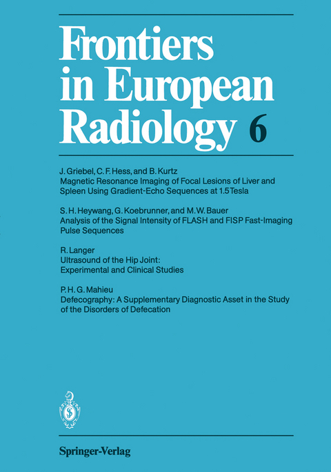
Frontiers in European Radiology
Seiten
2011
|
1. Softcover reprint of the original 1st ed. 1989
Springer Berlin (Verlag)
978-3-642-74235-4 (ISBN)
Springer Berlin (Verlag)
978-3-642-74235-4 (ISBN)
Congenital hip dysplasia and dislocation are common diseases of newborns and small infants, with frequently severe consequences if orthopaedic therapy is not initiated at an early stage. Therefore many clinicians have been looking for a simple method for the investigation of the hip joint in the early neonatal period. Up to 1980 the diagnosis of hip dysplasia could usually not be made before the 3rd month of life, by means of pelvic roentgenography. Only incomplete or complete unilateral dislocations were diagnosed in the neonatal age group. In 1980, however, Graf, an Austrian orthopaedic surgeon, began using ultrasound investigation ofthe hip joint in newborns and small infants in order to make an early diagnosis and to avoid radiation exposure. The intention of the present study was to compare ultrasound of the hip joint with other established diagnostic procedures and to establish whether it is suitable as a screening procedure in newborns. 2 Incidence of Congenital Hip Dysplasia and Dislocation In 1972 Barlow reported that 90 % of hips which are unstable at birth develop to normal joints spontaneously without any therapy. Visser (1984) thus suggested determining the percentage of hip dislocations after the 2nd - 3rd month of life so that children with spontaneous stabilisation would be excluded.
Magnetic Resonance Imaging of Focal Lesions of Liver and Spleen Using Gradient-Echo Sequences at 1.5 Tesla: A Comparison with Ultrasound and Sequential Computerized Tomography.- Analysis of the Signal Intensity of FLASH and FISP Fast-Imaging Pulse Sequences.- Ultrasound of the Hip Joint: Experimental and Clinical Studies.- Defecography: A Supplementary Diagnostic Asset in the Study of the Disorders of Defecation.
| Erscheint lt. Verlag | 6.12.2011 |
|---|---|
| Reihe/Serie | Frontiers in European Radiology |
| Co-Autor | J. Griebel, C.F. Hess, B. Kurtz, S.H. Heywang, G. Koebrunner, M.W. Bauer, R. Langer, P.H.G. Mahieu |
| Zusatzinfo | III, 87 p. |
| Verlagsort | Berlin |
| Sprache | englisch |
| Maße | 170 x 242 mm |
| Gewicht | 186 g |
| Themenwelt | Medizinische Fachgebiete ► Radiologie / Bildgebende Verfahren ► Sonographie / Echokardiographie |
| Technik | |
| Schlagworte | Diagnosis • hepatology • Hip • Imaging • Liver • Radiation • Radiology • Tomography • ultrasonography • Ultrasound |
| ISBN-10 | 3-642-74235-1 / 3642742351 |
| ISBN-13 | 978-3-642-74235-4 / 9783642742354 |
| Zustand | Neuware |
| Haben Sie eine Frage zum Produkt? |
Mehr entdecken
aus dem Bereich
aus dem Bereich
Begleitbuch für Sonografiekurse, Klinik und Praxis
Buch | Softcover (2023)
Urban & Fischer in Elsevier (Verlag)
27,00 €
Organbezogene Darstellung von Grund- und Aufbaukurs sowie …
Buch | Hardcover (2020)
Deutscher Ärzteverlag
99,99 €


