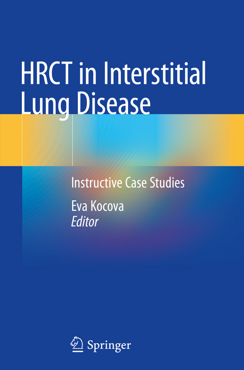
HRCT in Interstitial Lung Disease
Springer International Publishing (Verlag)
978-3-030-16317-4 (ISBN)
Eva Kocova graduated from Charles University, Prague (Faculty of Medicine in Hradec Kralove) in 2004. During her residency she worked in the Department of Radiology at Chrudim Hospital. After gaining her board certification in 2011, Dr. Kocova became a pulmonary radiology consultant at University Hospital Hradec Kralove. She is also an assistant professor of radiology at Charles University. In 2017 she completed her PhD, the topic of her thesis being Phenotypic assessment of patients with severe chronic obstructive pulmonary disease using HRCT of the lung. Dr. Kocova has published 27 scientific papers and two book chapters and has delivered more than 40 oral presentations, including 35 invited lectures. Her main fields of interest are pulmonary radiology and emergency radiology. She is a member of the Czech Radiological Society,the European Society of Radiology and the European Society of Thoracic Imaging.
General part: Introduction.- The role of multidisciplinary team in diagnosis and differential diagnosis of interstitial lung disease.- Pneumological basic in differential diagnosis of interstitial lung disease.- Radiological anatomy of the lung.- HRCT patterns.- Cases: Low attenuation patterns.- Linear opacities.- Nodulations.- High attenuation patterns.
| Erscheinungsdatum | 18.07.2020 |
|---|---|
| Zusatzinfo | XXII, 320 p. 338 illus., 79 illus. in color. |
| Verlagsort | Cham |
| Sprache | englisch |
| Maße | 155 x 235 mm |
| Gewicht | 584 g |
| Themenwelt | Medizinische Fachgebiete ► Innere Medizin ► Pneumologie |
| Medizinische Fachgebiete ► Radiologie / Bildgebende Verfahren ► Radiologie | |
| Schlagworte | diagnostic radiology • High attenuation patterns • High-Resolution CT • HRCT Patterns • Interstitial lung disease • Linear opacities • Low attenuation patterns • Low attenuation patterns • Lymfangioleiomyomatosis • Nodulations • Non-specific Interstitial Pneumonia • Pulmonary Langerhans Cell Histiocytosis • Pulmonary Medicine |
| ISBN-10 | 3-030-16317-2 / 3030163172 |
| ISBN-13 | 978-3-030-16317-4 / 9783030163174 |
| Zustand | Neuware |
| Haben Sie eine Frage zum Produkt? |
aus dem Bereich


