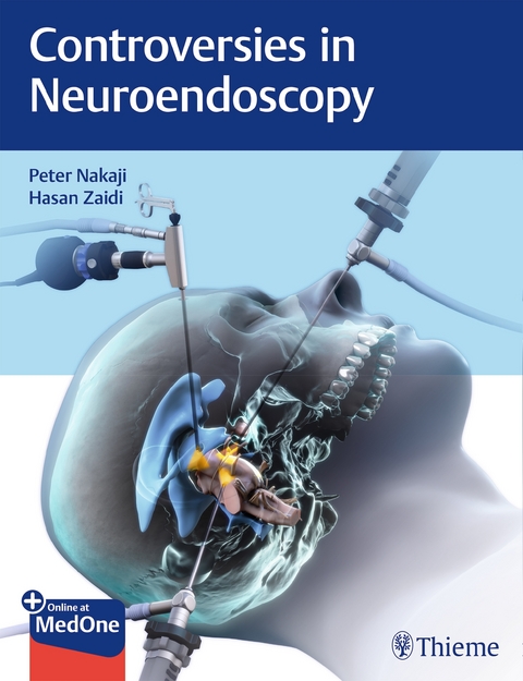
Controversies in Neuroendoscopy
Thieme Medical Publishers Inc (Verlag)
978-1-62623-353-9 (ISBN)
In the last two decades, neuroendoscopy has evolved from a fringe neurosurgical tool to an established subspecialty focusing on the treatment of diverse cranial and spinal diseases. Today, neuroendoscopic technology is widely used to treat supratentorial diseases, skull base pathologies, craniocervical diseases, and spinal pathologies.
Despite the expanded use of neuroendoscopy across several subspecialties, its benefits and disadvantages versus those of traditional microsurgical methods remain highly controversial. Contradictory evidence in the neurosurgical literature adds to the surgical decision-making challenges for veteran and novice practitioners alike.
»Controversies in Neuroendoscopy« by Peter Nakaji and Hasan Zaidi fills an unmet need for a book encompassing best practices, patient selection, and limitations and advantages of neuroendoscopic surgical approaches. Each case presents firsthand knowledge of internationally esteemed neurosurgeons, with a moderator, an endoscopic expert, and an expert in traditional microsurgical approaches. The unique discussion of neuroendoscopy versus microsurgery enables readers to compare the benefits and pitfalls of endoscopic and open microsurgical procedures for a wide range of conditions.
- In-depth comparative guidance on applications of the flexible endoscope, rigid endoscope, 3D endoscope, and high-definition 2D endoscope versus the microscope
- A full spectrum of neurological conditions across the age continuum with comparative approaches for skull base surgery, pituitary surgery, hydrocephalus, spinal surgery, peripheral nerve surgery, and arachnoid cyst fenestration
- Radiological imaging and intraoperative photographs enhance cases and provide precise, insightful technical guidance
- High-quality color illustrations from the skilled medical illustrators at Barrow Neurological Institute reinforce key points and surgical techniques
Neurosurgery residents, neurosurgeons, and spine surgeons will all benefit from reading this remarkable book cover to cover, and they will no doubt use it as a go-to resource for specific cases.
This book includes complimentary access to a digital copy on https://medone.thieme.com.
I History and Evolution of Neuroendoscopy
1. History and Evolution of Neuroendoscopy
II Skull Base Surgery
2. Microscopic versus Endoscopic Nasal Morbidity and Quality of Life after Skull Base Surgery
A. Pituitary Tumor Surgery
3. Pituitary Tumor Surgery: Endoscope versus Microscope-Moderator
4. Pituitary Tumor Surgery: Endoscope versus Microscope-Microscope
5. Pituitary Tumor Surgery: Endoscope versus Microscope-Endoscope
6. Endoscopic Approach to the Infratemporal Fossa
B. Anterior Skull Base Tumors
7. Anterior Skull Base Tumors-Microscope
8. Anterior Skull Base Tumors-Endoscope
C. Craniovertebral Junction
9. Endoscopic versus Microscopic Approach to the Craniovertebral Junction-Microscope
10. Craniovertebral Junction-Endoscope
III Paraventricular Lesions
11. Endoscopic Third Ventriculostomy versus Ventriculoperitoneal Shunting
D. Management of Colloid Cysts
12. Management of Colloid Cysts: Moderator
13. Management of Colloid Cysts: Microscope
14. Management of Colloid Cysts: Endoscope
15. Intracranial Arachnoid Cyst Fenestration versus Shunting
16. Endoscopic Aqueductal Stenting
IV Neurovascular
E. Decompression of Cranial Nerves
17. Decompression of Cranial Nerves: Microscope
18. Decompression of Cranial Nerves: Endoscope
F. Clipping of Cerebral Aneurysms
19. Clipping of Cerebral Aneurysms: Moderator
20. Clipping of Cerebral Aneurysms: Microscope and Endoscope
G. Evacuation of Intraparenchymal Hemorrhage
21. Evacuation of Intraparenchymal Hemorrhage: Moderator
22. Evacuation of Intraparenchymal Hemorrhage: Microscope
23. Evacuation of Intraparenchymal Hemorrhage: Endoscope
H. Approaches to Brainstem Cavernous Malformations
24. Approaches to Brainstem Cavernous Malformations: Microscope
25. Approaches to Brainstem Cavernous Malformations: Endoscope
V Tumors
26. Open versus Endoscopic Supracerebellar Infratentorial Approaches
I. Intraparenchymal Brain Tumors
27. Intraparenchymal Brain Tumors: Moderator
28. Intraparenchymal Brain Tumors: Microscope versus Endoscope
VI Pediatrics
29. Surgical Management of Hypothalamic Hamartomas
J. Craniosynostosis Surgery
30. Craniosynostosis Surgery: Moderator
31. Craniosynostosis Surgery: Microscope and Endoscope
VII Spine and Peripheral Nerves
K. Cervical Diskectomy/Foraminotomy
32. Cervical Diskectomy/Foraminotomy: Moderator
33. Cervical Diskectomy/Foraminotomy: Microscope
34. Cervical Diskectomy/Foraminotomy: Endoscope
L. Thoracic Diskectomy
35. Thoracic Diskectomy: Moderator
36. Thoracic Diskectomy: Microscope and Endoscopic
37. Spinal Lumbar Diskectomy: Open versus Endoscopic
M. Endoscopic versus Open Carpal Tunnel Release
38. Endoscopic versus Open Carpal Tunnel Release: Moderator
39. Endoscopic versus Open Carpal Tunnel Release: Endoscope
VIII Technology
40. 3D Endoscope versus High-Definition 2D Endoscope
41. Flexible versus Rigid Neuroendoscopy
42. Endoscopic Port Surgery: Advantages and Disadvantages
43. Future of Neuroendoscopy
| Erscheinungsdatum | 20.12.2017 |
|---|---|
| Zusatzinfo | 184 Abbildungen |
| Verlagsort | New York |
| Sprache | englisch |
| Maße | 216 x 279 mm |
| Gewicht | 1 g |
| Einbandart | gebunden |
| Themenwelt | Medizinische Fachgebiete ► Chirurgie ► Neurochirurgie |
| Medizin / Pharmazie ► Medizinische Fachgebiete ► HNO-Heilkunde | |
| Medizin / Pharmazie ► Medizinische Fachgebiete ► Orthopädie | |
| Schlagworte | controversies • Endoskopie • Nakaji • Neuroendoscopy • Neurologie • neurosurgery |
| ISBN-10 | 1-62623-353-5 / 1626233535 |
| ISBN-13 | 978-1-62623-353-9 / 9781626233539 |
| Zustand | Neuware |
| Haben Sie eine Frage zum Produkt? |
aus dem Bereich


