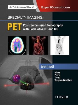
Specialty Imaging: PET
Elsevier - Health Sciences Division (Verlag)
978-0-323-52484-1 (ISBN)
Features 1,600 high-quality images with captions and annotations for interpretive guidance, with illustrations including PET, with correlative CT and MR images depicting radiologic imaging findings
Presents all diagnoses consistently, using a highly templated format with bulleted text for quick, easy reference
Includes chapters in expert interpretation, artifacts, and common pitfalls
Provides a wide range of essential information such as oncologic PET diagnoses with staging tables and reporting tips; cardiac PET indications including stress tests, cardiac viability, and sarcoidosis; CNS PET indications including dementia, epilepsy, and oncology; and educational, illustrated PET cases including correlative CT and MR
Covers PET physics and instrumentation and current clinical and emerging PET radiotracers in table format
Ideal for clinicians who care for cancer patients (nuclear medicine radiologists, radiation oncologists, oncologists, oncology surgeons, and trainees in nuclear medicine and oncology), as well as those who interpret PET for a wide variety of indications
Expert ConsultT eBook version included with purchase. This enhanced eBook experience allows you to search all of the text, figures, Q&As, and references from the book on a variety of devices.
Paige Bennett is a professor of nuclear medicine and molecular imaging with the Department of Radiology at Wake Forest School of Medicine in Winston-Salem, North Carolina
PET IMAGING: THE BASICS
Introduction to PET Imaging
Overview
PET Interpretation
Approach to Interpretation
Artifacts and Pitfalls
PET Protocols
Whole Body Protocols
Central Nervous System Protocols
Cardiac Protocols
Physics and Radiopharmaceuticals
Physics and Quality Control
Physics and Instrumentation
Radiopharmaceuticals
Clinical Radiopharmaceuticals
Radiopharmaceuticals in Development
PET IMAGING: CARDIAC
Myocardial Ischemia
Myocardial Viability
Cardiomyopathy
Acute Cardiac Infection and Inflammation
PET IMAGING: CENTRAL NERVOUS SYSTEM
Dementia
Alzheimer Disease
Frontotemporal Dementia
Corticobasal Dementia
Lewy Body Dementia
Posterior Cortical Atrophy
Multi-Infarct Dementia
Behavioral and Movement Disorders
Epilepsy
Intractable Seizure Disorder
CNS Infection and Inflammation
Cerebral Abscess and Encephalitis
Cerebral Toxoplasmosis
PET IMAGING: SOFT TISSUE INFECTION AND INFLAMMATION
Abdominal Infection and Inflammatory Disease
Pulmonary Infection and Inflammation
Granulomatous Disease
Fever of Unknown Origin
Vascular Infection and Inflammation
Large Vessel Vasculitis
PET IMAGING: MUSCULOSKELETAL
Pediatric Nonaccidental Trauma
Benign Bone Neoplasms
Bone and Joint Infection
PET IMAGING: ONCOLOGY
Adrenal
Adrenal Carcinoma
Breast Oncology
Primary Breast Cancer
Breast Cancer Staging
Central Nervous System Oncology
Primary Brain Malignancy
Brain Metastases
Cutaneous Oncology
Melanoma
Squamous Cell Carcinoma
Merkel Cell Carcinoma
Gastrointestinal Oncology
Esophageal Cancer
Gastric Cancer and Gastrointestinal Stromal Tumor
Anal Carcinoma
Peritoneal Carcinoma and Mesothelioma
Genitourinary Oncology
Uterine and Endometrial Cancers
Ovarian Cancer
Cervical Cancer
Vulvar and Vaginal Cancer
Prostate Cancer
Testicular Cancer
Renal Cell Carcinoma
Transitional Cell Carcinoma
Head and Neck Cancer
Squamous Cell Carcinoma
Parotid and Salivary Gland Carcinoma
Hepatobiliary Oncology
Hepatocellular Carcinoma
Adrenal Oncology
Pancreatic Oncology
Pancreatic Adenocarcinoma
Pancreatic Islet Cell Tumor/Neuroendocrine
Thoracic Oncology
Pulmonary Carcinoid Tumor
Lung Adenocarcinoma
Malignant Pleural Mesothelioma
Small Cell Lung Cancer
Solitary Pulmonary Nodule
Thymoma and Thymic Carcinoma
Thyroid and Parathyroid Carcinoma
Anaplastic Thyroid Carcinoma
Papillary and Follicular Thyroid Cancer
Medullary Thyroid Carcinoma
Parathyroid Carcinoma
Lymphoma
Hodgkin Lymphoma
Non-Hodgkin Lymphoma
Musculoskeletal Oncology
Primary Bone Tumors
Multiple Myeloma
Sarcomas
Neuroendocrine Oncology
Abdominal Carcinoid Tumors
Pancreatic Neuroendocrine Tumors
Pheochromocytoma and Paraganglioma
Pediatric Oncology
Neuroblastoma
Ewing Sarcoma
Osteosarcoma
Pediatric Lymphoma
PET: EMERGING CLINICAL APPLICATIONS
| Erscheinungsdatum | 02.01.2018 |
|---|---|
| Reihe/Serie | Specialty Imaging |
| Verlagsort | Philadelphia |
| Sprache | englisch |
| Maße | 216 x 276 mm |
| Gewicht | 1340 g |
| Themenwelt | Medizin / Pharmazie ► Medizinische Fachgebiete ► Onkologie |
| Medizinische Fachgebiete ► Radiologie / Bildgebende Verfahren ► Nuklearmedizin | |
| Medizinische Fachgebiete ► Radiologie / Bildgebende Verfahren ► Radiologie | |
| ISBN-10 | 0-323-52484-2 / 0323524842 |
| ISBN-13 | 978-0-323-52484-1 / 9780323524841 |
| Zustand | Neuware |
| Haben Sie eine Frage zum Produkt? |
aus dem Bereich


