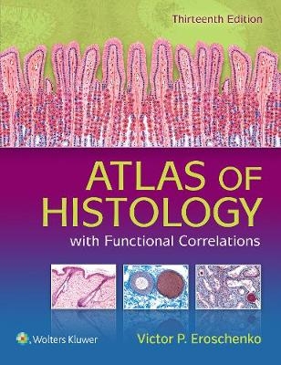
Atlas of Histology with Functional Correlations
Seiten
2017
|
Thirteenth, North American Edition
Lippincott Williams and Wilkins (Verlag)
978-1-4963-1676-9 (ISBN)
Lippincott Williams and Wilkins (Verlag)
978-1-4963-1676-9 (ISBN)
- Titel erscheint in neuer Auflage
- Artikel merken
Master histology with idealized and actual photomicrography!
This thirteenth edition of Atlas of Histology with Functional Correlations (formerly diFiore’s ) provides a rich understanding of the basic histology concepts that medical and allied health students need to know. Realistic, full-color illustrations as well as actual photomicrographs of histologic structures are complemented by concise discussions of their most important functional correlations.
Illustrated histology images show the idealized view, while photomicrographs provide the actual view to help students hone their skills in identifying structures.
New and improved layout helps students connect the morphology of a structure with its function.
Updated and expanded Functional Correlations boxes integrated throughout chapters reflect new scientific information and interpretations.
NEW photomicrographs and electron micrographs provide views of microanatomy as experienced in practice.
Bulleted Chapter Summaries distill the most essential knowledge for rapid review.
NEW Additional Histologic Images sections round out each chapter with supplemental photomicrographs and electron micrographs.
NEW Chapter Review Questions allow students to assess their comprehension of each chapter with 375 questions and answers in the book and 250 more online in an Interactive Question Bank.
| Erscheinungsdatum | 19.03.2017 |
|---|---|
| Zusatzinfo | 362 |
| Verlagsort | Philadelphia |
| Sprache | englisch |
| Maße | 213 x 276 mm |
| Gewicht | 1701 g |
| Themenwelt | Studium ► 1. Studienabschnitt (Vorklinik) ► Histologie / Embryologie |
| ISBN-10 | 1-4963-1676-2 / 1496316762 |
| ISBN-13 | 978-1-4963-1676-9 / 9781496316769 |
| Zustand | Neuware |
| Haben Sie eine Frage zum Produkt? |
Mehr entdecken
aus dem Bereich
aus dem Bereich
Zytologie, Histologie und mikroskopische Anatomie
Buch | Hardcover (2022)
Urban & Fischer in Elsevier (Verlag)
54,00 €
Gewebelehre, Organlehre
Buch | Spiralbindung (2024)
Urban & Fischer in Elsevier (Verlag)
25,00 €


