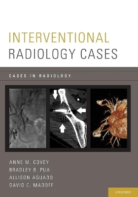
Interventional Radiology Cases
Oxford University Press Inc (Verlag)
978-0-19-933127-7 (ISBN)
In 104 cases featuring over 500, high-quality images, Interventional Radiology Cases is a thorough and accessible review of the interventional procedures that radiology residents are expected to be familiar with upon completion of residency and general radiologists need to know for recertification examinations. The cases present both benign and malignant conditions and all pertinent imaging modalities incorporated including: CT, MR, PET, fluoroscopy, and ultrasound. Part of the Cases in Radiology series, this book follows the easy-to-use format of question and answer in which the patient history is provided on the first page of the case, and radiologic findings, differential diagnosis, teaching points, next steps in management, and suggestions for furthering reading are revealed on the following page.
AMC: Associate Professor of Radiology, Division of Interventional Radiology, Memorial Sloan Kettering Cancer Center, Weill Cornell Medical College, New York, New York. DM: Chief of Division of Interventional Radiology, Professor of Radiology, New York Presbyterian Hospital, Weill Cornell Medical College, New York, New York. AA: Assistant Professor of Radiology, Division of Interventional Radiology, Memorial Sloan Kettering Cancer Center, Weill Cornell Medical College, New York, New York. BP: Assistant Professor of Radiology, Division of Interventional Radiology, New York Presbyterian Hospital, Weill Cornell Medical College, New York, New York.
Case List ; 1. Mediport ; 2. Translumbar placement of implantable venous access device (IVAD) ; 3. Left Superior Vena Cava ; 4. Port Catheter Dislodged From Reservoir ; 5. Calcified Fibrin Sheath ; 6. Lymphangiogram and Thoracic Duct Embolization ; 7. Post Biopsy Pneumothorax ; 8. Mediastinal Biopsy Utilizing Separation Techniques ; 9. Right Coronary Artery Aneurysm ; 10. PET Guided Biopsy of Left Iliac Bone Metastasis ; 11. Adrenal Biopsy ; 12. Transcaval Pancreatic Biopsy ; 13. Autoimmune pancreatitis (AIP) with Biliary Tract Involvement ; 14. Transjugular Liver Biopsy, Elevated Hepatic Venous Pressure Gradient ; 15. Renal PSA ; 16. Transperineal MR Guided Biopsy ; 17. Celiac Plexus Neurolysis ; 18. Suspicious Liver Lesions for Biopsy ; 19. Suspicious Thyroid Nodule for Biopsy ; 20. Placement of fiducial markers to facilitate image-guided radiation therapy ; 21. Incomplete Coverage Of Embolized Hcc Treated With Microwave Ablation ; 22. Lung Ablation ; 23. Renal Cell Carcinoma treated with Cryoablation ; 24. Metastatic osseous lesion treated with cryoablation for pain palliation ; 25. Osteoid Osteoma ; 26. Hypertensive Crisis during Adrenal Radiofrequency Ablation ; 27. Occluded Celiac Artery With Retrograde Flow Through Gastroduodenal Artery ; 28. Liver Abscess After Embolization in a Patient S/P Pancreaticoduodenectomy ; 29. Transarterial Embolization with Y-90 Particles ; 30. Hepatic Artery Aneurysm ; 31. Pre Operative Embolization Of Renal Cell Carcinoma Bone Metastasis From Right Radial Artery Approach ; 32. Bronchial Artery Embolization ; 33. Pulmonary Arteriovenous Malformation (PAVM) ; 34. Pelvic Congestion Syndrome Treated with Embolization ; 35. Ruptured Renal Angiomyolipoma ; 36. Liver Trauma ; 37. Splenic Trauma ; 38. Drainage of Abdominal Abscess Complicated by Inferior Epigastric Artery Pseudoaneurysm ; 39. Upper GI Bleed ; 40. Lower GI Bleed ; 41. Juvenile Nasal Angiofibroma ; 42. Male Varicocele Embolized with Coils and Sclerosant ; 43. Partial Splenic Embolization to Treat Thrombocytopenia ; 44. Retrieval of non-target coil from the right hepatic artery during emboliztion of the gastroduodenal artery for gastrointestinal hemorrhage ; 45. Hydronephrosis from obstructing mass failing ureteral stent treatment ; 46. Ureteral-right iliac artery fistula ; 47. Conversion of Nephroureterostomy Catheters to Retrograde Nephrostomy Catheters ; 48. Ureteral Stent ; 49. Routine Transurethral Exchange of Ureteral Stent ; 50. Occluded Ureteral Stent ; 51. Ureterorectal Fistula ; 52. Peritoneo-venous Shunt Placement for Chylous Ascites ; 53. Percutaneous radiological jejunostomy catheter placement ; 54. Percutaneous Cecostomy with Retention Sutures Complicated by Fasciitis ; 55. Percutaneous Pull-Through Gastrostomy Complicated by Stomal Tumor Seeding ; 56. Tunneled Drainage Placement for Relief of Recurrent Ascites ; 57. Internal/External Biliary Drain ; 58. Persistent Leak Into Intrahepatic Biloma ; 59. High Bile Duct Obstruction with Isolation of the Right and Left Bile Ducts ; 60. Biliary Stent Complicated By Cholecystitis ; 61. Occluded Wallstent complicated by cholangitis and cholangitic abscesses ; 62. Varicose Veins - Greater Saphenous Vein Reflux ; 63. Transjugular Intrahepatic Portosystemic Shunt (TIPS) ; 64. Balloon-Occluded Retrograde Transvenous Obliteration of Gastric Varices ; 65. Adrenal Vein Sampling ; 66. Superior Vena Cava Syndrome ; 67. Paget Schroetter ; 68. Sclerosis of Lymphatic Malformation ; 69. May Thurner Syndrome ; 70. Inferior Vena Cava (IVC) Filter Placement ; 71. Inferior Vena Cava (IVC) Filter Removal ; 72. Inferior Vena Cava (IVC) Stent ; 73. Lower Extremity Lysis ; 74. Acute Pulmonary Embolism ; 75. Duplicated Inferior Vena Cava (IVC) ; 76. Type B Aortic Dissection and Fenestration ; 77. Endoleak ; 78. Traumatic Psa Carotid ; 79. Acute Traumatic Aortic Injury ; 80. Renal Artery Stenosis ; 81. Hepatic Artery Stricture After S/P Transplant ; 82. Femoral Artery Pseudoaneurysm ; 83. Acute Superior Mesenteric Artery Embolism ; 84. Uterine Fibroid Embolization (UFE) ; 85. Popliteal Entrapment ; 86. Portal Vein Embolization to Induce Contralateral Hepatic Hypertrophy ; 87. Takayasu's Arteritis with Ascending Aortic Aneurysm ; 88. Leriche Syndrome ; 89. Thoracic Outlet Syndrome and Subclavian Artery Aneurysm ; 90. Variant Hepatic Arterial Anatomy: Replaced Right Hepatic Artery ; 91. Venous Malformation Sclerosis ; 92. Subclavian Steal ; 93. Arteriovenous Malformation (AVM) ; 94. Ruptured Appendix ; 95. Pelvic Abscess - Transgluteal Drainage ; 96. Transrectal Aspiration of Prostatic Abscess ; 97. Chemical Sclerosis of Traumatic Lymphocele ; 98. Small Bowel Fistula ; 99. Loculated Effusion Treated with Tunneled Pleural Catheter ; 100. Anastomotic Leak ; 101. Esophageal Leak, S/P Covered Stent and Surgical Chest Tube placement with Persistent Locaulated Infected Collection (Empyema) ; 102. Pericardial Drain ; 103. Needle tract Seeding Post Biopsy of Hepatocellular Carcinoma ; 104. Left Kyphoplasty and Right Vertebroplasty
| Erscheint lt. Verlag | 12.3.2015 |
|---|---|
| Reihe/Serie | Cases In Radiology |
| Zusatzinfo | 565 illustrations |
| Verlagsort | New York |
| Sprache | englisch |
| Maße | 178 x 251 mm |
| Gewicht | 816 g |
| Themenwelt | Medizin / Pharmazie ► Medizinische Fachgebiete ► Chirurgie |
| Medizinische Fachgebiete ► Radiologie / Bildgebende Verfahren ► Radiologie | |
| ISBN-10 | 0-19-933127-8 / 0199331278 |
| ISBN-13 | 978-0-19-933127-7 / 9780199331277 |
| Zustand | Neuware |
| Haben Sie eine Frage zum Produkt? |
aus dem Bereich


