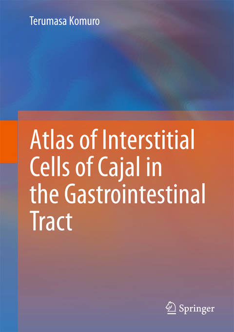
Atlas of Interstitial Cells of Cajal in the Gastrointestinal Tract
Seiten
This atlas reviews the distribution and morphological features of interstitial cells of Cajal, key to understanding the regulatory mechanism of gastrointestinal motility. Includes three-dimensional reconstruction of confocal images, and electron micrographs.
This atlas will illustrate the distribution and morphological features of interstitial cells of Cajal (ICC) which are the key cells to understanding of the regulatory mechanism of gastrointestinal motility, since ICC act as both pacemaker and as intermediates in neural transmission, and since ICC show specific distribution patterns depending on their anatomical positions. All subtypes of ICC located in the different tissue layers and different levels of the gastrointestinal tract will be revealed by immunohistochemistry for Kit receptors and nerves by using mainly whole-mount stretch preparation of the guinea-pig tissues. Three-dimensional reconstruction of confocal images will particularly help the readers to understand the peculiar arrangement of ICC networks in situ and the correlation between ICC and nerves. Electron micrographs will help illustrate the characteristic features of ICC and their ultrastructural differences from fibroblasts, smooth muscles and other interstitial cells.
This atlas will illustrate the distribution and morphological features of interstitial cells of Cajal (ICC) which are the key cells to understanding of the regulatory mechanism of gastrointestinal motility, since ICC act as both pacemaker and as intermediates in neural transmission, and since ICC show specific distribution patterns depending on their anatomical positions. All subtypes of ICC located in the different tissue layers and different levels of the gastrointestinal tract will be revealed by immunohistochemistry for Kit receptors and nerves by using mainly whole-mount stretch preparation of the guinea-pig tissues. Three-dimensional reconstruction of confocal images will particularly help the readers to understand the peculiar arrangement of ICC networks in situ and the correlation between ICC and nerves. Electron micrographs will help illustrate the characteristic features of ICC and their ultrastructural differences from fibroblasts, smooth muscles and other interstitial cells.
1 Introduction; 2 Stomach; 3 Small intestine; 4 Duodenum; 5 Colon; 6 Caecum; 7 Ileocaecal Junction; 8 ICC found in the submucosal layer; 9 Ultrastructural demonstration of ICC; 10 Signal-pathways between the nerves and muscles not by means of ICC; 11 Issues for future studies
| Zusatzinfo | XII, 134 p. |
|---|---|
| Verlagsort | Dordrecht |
| Sprache | englisch |
| Maße | 178 x 254 mm |
| Themenwelt | Medizinische Fachgebiete ► Innere Medizin ► Gastroenterologie |
| Medizin / Pharmazie ► Studium | |
| Schlagworte | c-kit • Gastrointestinaltrakt • GI Tract • ICC • Morphology • Smooth musculature |
| ISBN-10 | 94-007-2916-2 / 9400729162 |
| ISBN-13 | 978-94-007-2916-2 / 9789400729162 |
| Zustand | Neuware |
| Haben Sie eine Frage zum Produkt? |
Mehr entdecken
aus dem Bereich
aus dem Bereich
Buch | Softcover (2024)
Urban & Fischer in Elsevier (Verlag)
59,00 €
