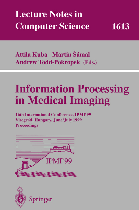
Information Processing in Medical Imaging
Springer Berlin (Verlag)
978-3-540-66167-2 (ISBN)
New Imaging Techniques.- Analytical Study of Bioelasticity Ultrasound Systems.- MEG Source Imaging Using Multipolar Expansions.- Binary Tomography for Triplane Cardiography.- Real Time 3D Brain Shift Compensation.- 3D Ultrasound and PET.- Computer Assisted Human Follicle Analysis for Fertility Prospects with 3D Ultrasound.- Volume Measurement in Sequential Freehand 3-D Ultrasound.- Automated Identification and Measurement of Objects via Populations of Medial Primitives, with Application to Real Time 3D Echocardiography.- Continuous Time Dynamic PET Imaging Using List Mode Data.- Segmentation.- Hybrid Geometric Active Models for Shape Recovery in Medical Images.- Co-dimension 2 Geodesic Active Contours for MRA Segmentation.- An Adaptive Fuzzy Segmentation Algorithm for Three-Dimensional Magnetic Resonance Images.- Automatic Detection and Segmentation of Evolving Processes in 3D Medical Images: Application to Multiple Sclerosis.- Image Analysis of the Brain Cortex.- Registration of Cortical Anatomical Structures via Robust 3D Point Matching.- Hierarchical Matching of Cortical Features for Deformable Brain Image Registration.- Using Local Geometry to Build 3D Sulcal Models.- ANIMAL+INSECT: Improved Cortical Structure Segmentation.- Registration.- Consistent Linear-Elastic Transformations for Image Matching.- Non-linear Registration with the Variable Viscosity Fluid Algorithm.- Approximating Thin-Plate Splines for Elastic Registration: Integration of Landmark Errors and Orientation Attributes.- A Hierarchical Feature Based Deformation Model Applied to 4D Cardiac SPECT Data.- Feature Detection and Modelling.- Local Orientation Distribution as a Function of Spatial Scale for Detection of Masses in Mammograms.- Physiologically Oriented Models of the Hemodynamic Response in Functional MRI.- 3D Graph Description of the Intracerebral Vasculature from Segmented MRA and Tests of Accuracy by Comparison with X-ray Angiograms.- A Unified Framework for Atlas Matching Using Active Appearance Models.- Poster Session I.- An Integral Method for the Analysis of Wall Motion in Gated Myocardial SPECT Studies.- Enhanced Artery Visualization in Blood Pool MRA: Results in the Peripheral Vasculature.- Four-Dimensional LV Tissue Tracking from Tagged MRI with a 4D B-Spline Model.- Recovery of Soft Tissue Object Deformation from 3D Image Sequences Using Biomechanical Models.- Forward Deformation of PET Volumes Using Non-uniform Elastic Material Constraints.- Brain Morphometry by Distance Measurement in a Non-Euclidean, Curvilinear Space.- Learning Shape Models from Examples Using Automatic Shape Clustering and Procrustes Analysis.- A Framework for Automated Landmark Generation for Automated 3D Statistical Model Construction.- Statistical Shape Analysis Using Fixed Topology Skeletons: Corpus Callosum Study.- Model Generation from Multiple Volumes Using Constrained Elastic SurfaceNets.- An Intelligent Interactive Segmentation Method for the Joint Space in Osteoarthritic Ankles.- Anatomical Modeling with Fuzzy Implicit Surfaces: Application to Automated Localization of the Heart and Lungs in Thoracic MR Images.- Detection of the Central Mass of Spiculated Lesions - Signature Normalisation and Model Data Aspects.- Noise Estimation and Measures for Detection of Clustered Microcalcifications.- Poster Session II.- Measuring the Spatial Homogeneity in Corneal Endotheliums by Means of a Randomization Test.- The Assessment of Chronic Liver Diseases by Sonography.- Automatic Computation of Brain and Cerebellum Volumes in Normal Subjects and Chronic Alcoholics.-Reconstruction from Slow Rotation Dynamic SPECT Using a Factor Model.- Spectral Factor Analysis for Multi-isotope Imaging in Nuclear Medicine.- Structural Group Analysis of Functional Maps.- Incorporating an Image Distortion Model in Non-rigid Alignment of EPI with Conventional MRI.- The Distribution of Target Registration Error in Rigid-Body, Point-Based Registration.- A Fast Mutual Information Method for Multi-modal Registration.- Voxel Similarity Measures for 3D Serial MR Brain Image Registration.- Radial Basis Function Interpolation for Freehand 3D Ultrasound.- Nonlinear Smoothing of MR Images Using Approximate Entropy - A Local Measure of Signal Intensity Irregularity.- New Variants of a Method of MRI Scale Normalization.- Method for Estimating the Intensity Mapping between MRI Images.
| Erscheint lt. Verlag | 16.6.1999 |
|---|---|
| Reihe/Serie | Lecture Notes in Computer Science |
| Zusatzinfo | XVII, 396 p. |
| Verlagsort | Berlin |
| Sprache | englisch |
| Gewicht | 750 g |
| Themenwelt | Informatik ► Grafik / Design ► Digitale Bildverarbeitung |
| Schlagworte | 3D • Bildgebendes Verfahren • Bildverarbeitung • Biologische Medizin • Biologische Medizin / Biomedizin • Hardcover, Softcover / Informatik, EDV/Informatik • HC/Informatik, EDV/Informatik • Image Analysis • Image Processing • Image Sequences • Imaging techniques • Informationsverarbeitung • Medical Imaging • Modeling • Mustererkennung • Segmentation • Ultrasound |
| ISBN-10 | 3-540-66167-0 / 3540661670 |
| ISBN-13 | 978-3-540-66167-2 / 9783540661672 |
| Zustand | Neuware |
| Haben Sie eine Frage zum Produkt? |
aus dem Bereich


